
Cell Biology Protocols
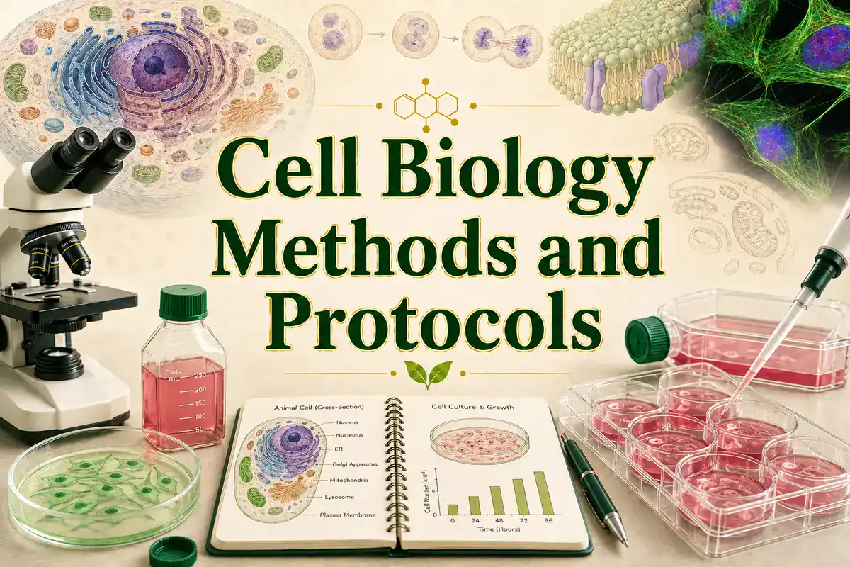
Cell biology protocols are practical laboratory methods used to grow, maintain, observe, count, treat, stain, image, and analyze cells. These methods help researchers study cell structure, cell division, cell signaling, cell death, cell migration, cell differentiation, organelles, membranes, cytoskeleton, cell-cell interactions, and responses to genes, drugs, pathogens, or environmental conditions.
This page is a guide to major cell biology methods and protocols used in research labs, teaching labs, biotechnology, immunology, cancer biology, developmental biology, neuroscience, microbiology, molecular biology, and biomedical science. It explains what each method is for, what a good protocol should include, where mistakes usually happen, and which trusted resources can help you go deeper.
A useful protocol is more than a list of steps. It should explain the purpose of the method, sample type, culture conditions, controls, reagents, equipment, timing, quality checks, data readout, safety issues, troubleshooting, and limitations. Nature Protocols describes strong protocols as including reagents, equipment, timing, procedures, design advice, limitations, troubleshooting, data analysis, and result interpretation.
What Are Cell Biology Protocols?
Cell biology protocols are written workflows for studying cells under defined conditions. They may be used to maintain a mammalian cell line, passage adherent cells, freeze cells, thaw cells, count live cells, measure viability, introduce DNA or RNA, stain organelles, detect proteins, analyze cell cycle, quantify apoptosis, or image cell behavior under a microscope.
The method is the scientific technique. The protocol is the practical workflow for using that technique. For example, cell counting is a method for estimating cell number. A cell counting protocol describes the sample preparation, dilution, stain, counting device, calculation, viability rule, and how the result will be used for seeding, passaging, freezing, or assay setup.
Cell biology overlaps closely with cell biology, molecular biology, biochemistry, immunology, developmental biology, neuroscience, genetics, cancer biology, physiology, and biotechnology.
Cell Biology Protocols Guide
Use this page as a map for common cell biology lab methods and cell-based workflows.
- aseptic technique and contamination control
- cell culture and media preparation
- cell passaging and subculturing
- cryopreservation and cell thawing
- cell counting and viability testing
- cell seeding density and assay setup
- transfection and gene delivery
- cell viability and proliferation assays
- apoptosis and cell death assays
- immunofluorescence and cell staining
- fluorescence microscopy and live-cell imaging
- flow cytometry and cell sorting
- cell cycle analysis
- migration, invasion, and wound healing assays
- controls, troubleshooting, and quality checks
Core Cell Biology Methods at a Glance
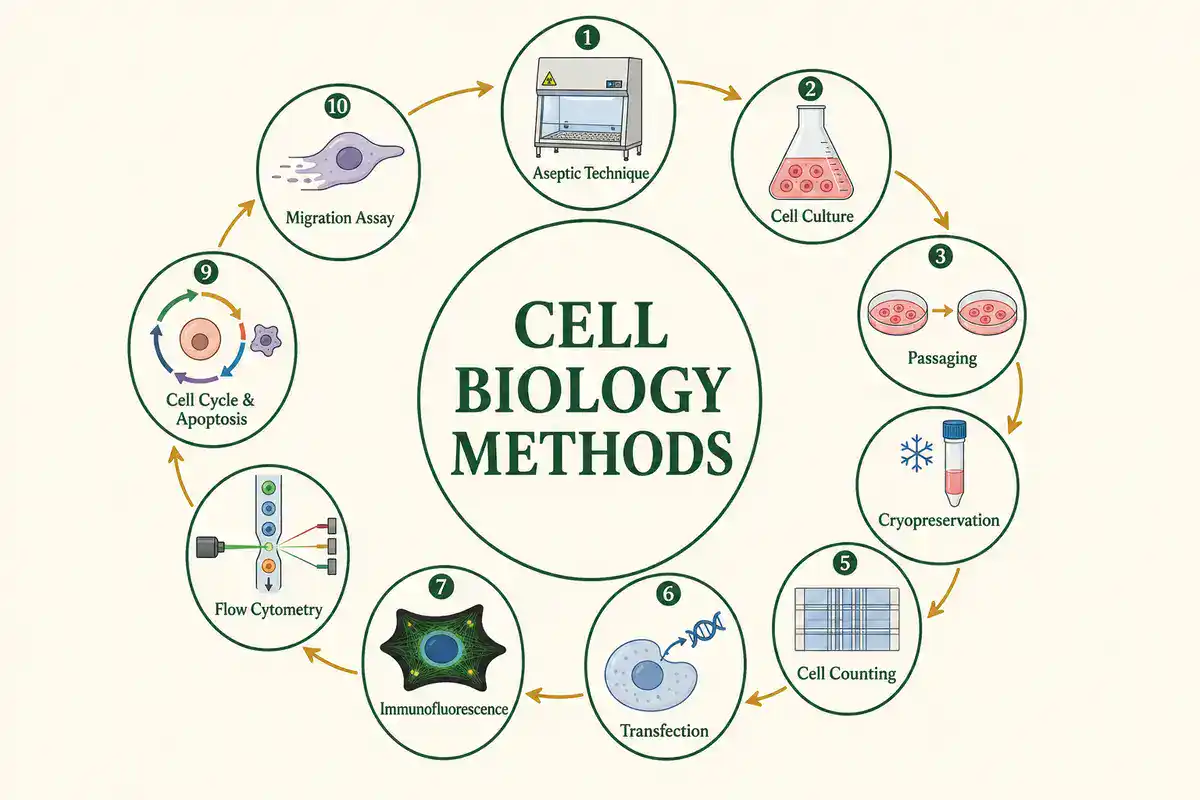
The table below gives a quick map of important cell biology methods and what each one helps measure or control.
| Method | Main Purpose | Common Readout |
|---|---|---|
| Cell culture | Grow and maintain cells under controlled conditions | Cell morphology, confluence, growth rate, viability, contamination status |
| Aseptic technique | Reduce contamination during cell handling | Sterile cultures, clean media, absence of visible microbial contamination |
| Passaging | Move growing cells into fresh vessels before overgrowth | Split ratio, passage number, confluence, recovery after subculture |
| Cryopreservation | Freeze cells for long-term storage | Post-thaw viability, recovery, morphology, growth after thawing |
| Cell counting | Estimate cell number for seeding, passaging, or assays | Cells/mL, total cells, viable cells, percent viability |
| Viability assay | Estimate live or metabolically active cells | Percent viability, relative signal, dose response, treatment effect |
| Transfection | Introduce DNA, RNA, or other molecules into cells | Reporter expression, knockdown, editing signal, transfection efficiency |
| Immunofluorescence | Detect proteins or structures in cells using antibodies and fluorescent labels | Fluorescent staining pattern, localization, intensity, colocalization |
| Flow cytometry | Measure physical and fluorescent properties of single cells in suspension | Cell populations, marker expression, scatter, fluorescence intensity, gating results |
| Migration assay | Measure cell movement across a surface or membrane | Scratch closure, migrated cell count, invasion index, migration rate |
For techniques that measure membrane activity, cell mechanics, and single molecule forces, see BioExplorer’s Biophysics Methods and Protocols guide.
Cell Culture and Aseptic Technique
Cell culture protocols describe how to grow cells outside the organism under controlled conditions. Cell culture may involve mammalian cells, insect cells, plant cells, primary cells, immortalized cell lines, stem cells, organoids, co-cultures, or microbial cells, depending on the experiment.
ATCC's animal cell culture guide covers media, subculturing, cryopreservation, and contamination for animal cells in culture. See ATCC Animal Cell Culture Guide. Thermo Fisher's cell culture basics handbook explains that aseptic technique is designed to create a barrier between microorganisms in the environment and sterile cell culture, using a sterile work area, good hygiene, sterile reagents, and sterile handling. See Gibco Cell Culture Basics Handbook.
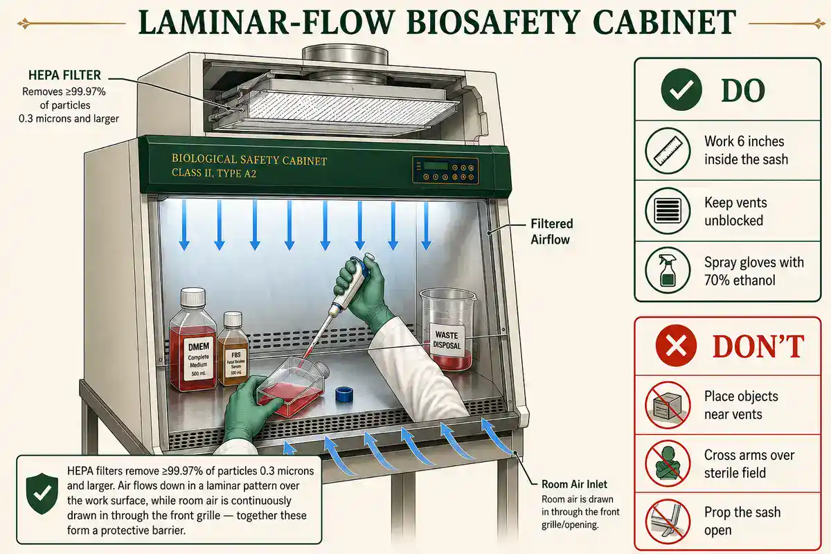
A useful cell culture protocol should clearly state the cell type, source, medium, serum or supplement conditions, culture vessel, temperature, CO2, humidity, passage range, seeding density, split ratio, medium-change schedule, contamination checks, and discard criteria. It should also state whether the cells are adherent, suspension, primary, immortalized, contact-inhibited, serum-dependent, or sensitive to passage number.
Cell culture is not just "keeping cells alive." The goal is to keep cells healthy, consistent, and appropriate for the experiment. Two flasks can contain the same cell line but behave differently if passage number, confluence, media age, serum lot, contamination history, or handling differ.
Where Do Stem Cells Come From?
Contamination Control and Cell Line Quality
Contamination is one of the most important quality problems in cell biology. Bacteria, fungi, yeast, viruses, cross-contaminating cell lines, and mycoplasma can change cell growth, morphology, gene expression, metabolism, signaling, assay response, and interpretation.
Mycoplasma contamination is especially difficult because it may not always make cultures look obviously cloudy. ATCC's animal cell culture guide describes direct and indirect approaches for detecting mycoplasma contamination, including culture methods and Hoechst staining. A practical cell culture review also discusses mycoplasma detection and emphasizes routine contamination awareness. See ATCC Animal Cell Culture Guide and A Beginner’s Guide to Cell Culture.
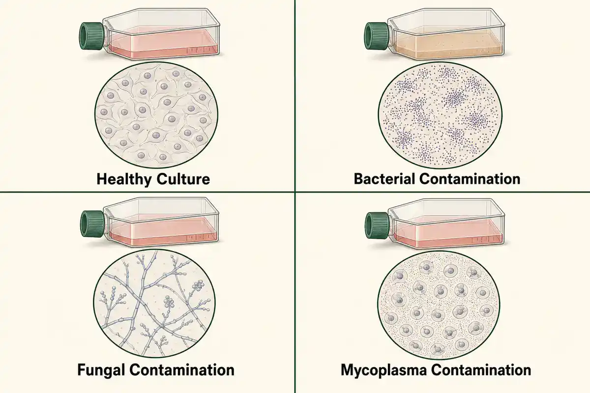
A strong cell biology protocol should include quality checkpoints. These may include cell morphology, viability, passage number, confluence, mycoplasma testing, authentication for established cell lines, contamination inspection, and records of thaw date, passage date, and treatment history.
For serious research, avoid relying on appearance alone. A culture can look acceptable under the microscope and still carry a hidden problem that affects results.
Passaging, Seeding Density and Cryopreservation
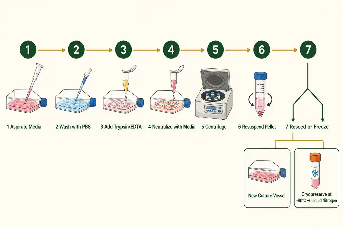
Passaging protocols, also called subculturing protocols, describe how cells are moved into fresh vessels before they overgrow, exhaust nutrients, change behavior, or lose experimental consistency. The details depend on whether the cells grow attached to a surface or in suspension.
A useful passaging protocol should state the target confluence, detachment method if relevant, wash conditions, dissociation reagent, neutralization step, centrifugation if used, resuspension medium, split ratio, seeding density, and passage number recording. For adherent cells, over-digestion during detachment can damage cells. For suspension cells, density and aggregation can matter more than surface detachment.
Cryopreservation protocols are used to store cells for future experiments. They should describe freezing medium, cryoprotectant, cell density, cooling method, storage temperature, vial labeling, recovery conditions, and post-thaw viability checks. A good freezing protocol protects the future experiment, not just the current stock.
The most useful cell culture records include cell line name, source, passage number, thaw date, split dates, medium, supplements, contamination checks, experimental treatments, and any deviations from the usual culture workflow.
Cell Counting and Viability Protocols
Cell counting protocols estimate how many cells are present in a sample. Counting is used before seeding plates, passaging cultures, freezing stocks, transfecting cells, running viability assays, preparing flow cytometry samples, and setting up treatment experiments.
Cell counting may use a hemocytometer, automated cell counter, image-based counter, flow cytometer, or plate reader-based proxy, depending on the experiment. A strong protocol should state how cells are mixed, diluted, stained, loaded, counted, excluded, and calculated. If viability is measured, the protocol should define what counts as live or dead.
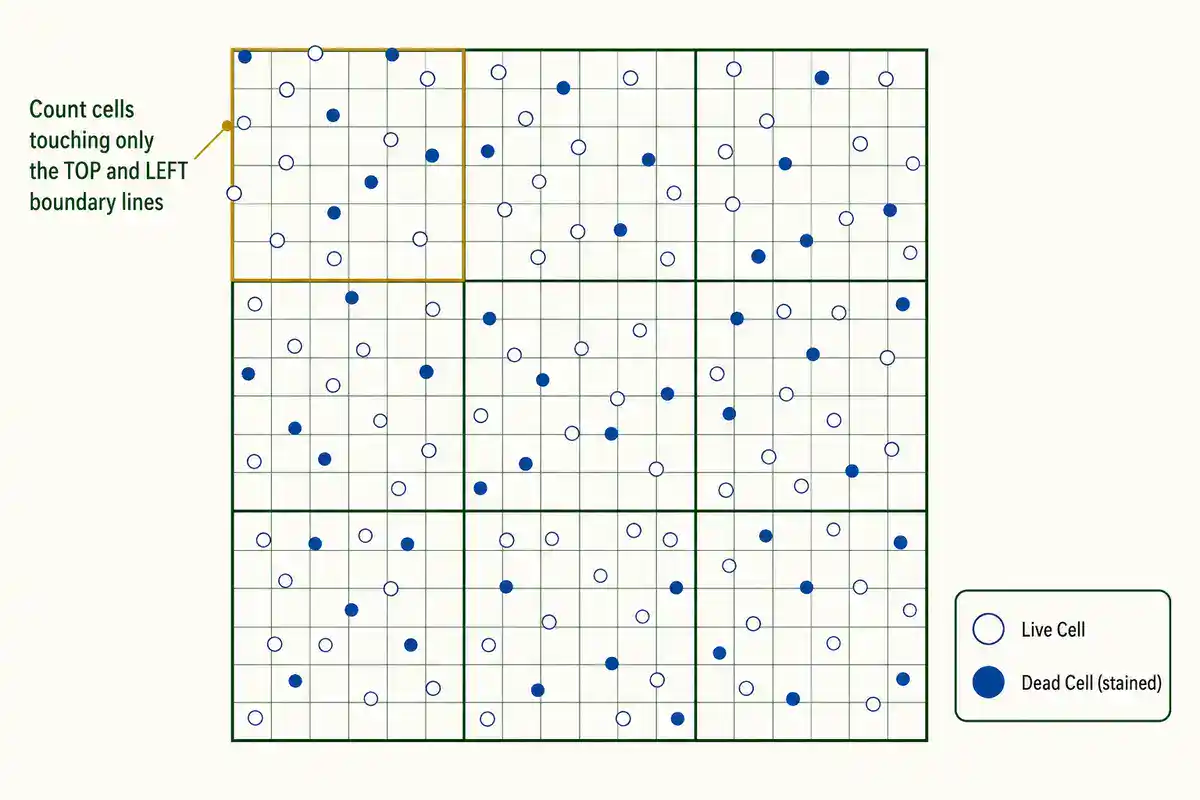
Common viability methods include dye exclusion, metabolic activity assays, fluorescent live-dead stains, apoptosis assays, and flow cytometry-based measurements. These methods are not interchangeable. A metabolic assay may indicate metabolic activity, not direct cell number. A dye exclusion assay may miss early stress responses. A flow assay may provide richer single-cell information but needs proper gating and controls.
Good cell counting and viability protocols should also include replicate strategy, acceptable viability range, treatment timing, dilution factor, instrument settings, and rules for handling clumps, debris, edge effects, and uneven cell suspensions.
Transfection and Gene Delivery Protocols
Transfection protocols introduce DNA, RNA, or other molecules into cells. They are used for gene expression, reporter assays, knockdown, CRISPR editing, protein localization, pathway analysis, and functional screening.
Transfection may use lipid-based reagents, polymers, electroporation, calcium phosphate, viral vectors, microinjection, or other delivery methods. The right approach depends on cell type, molecule type, toxicity, expression timeline, transfection efficiency, and downstream readout.
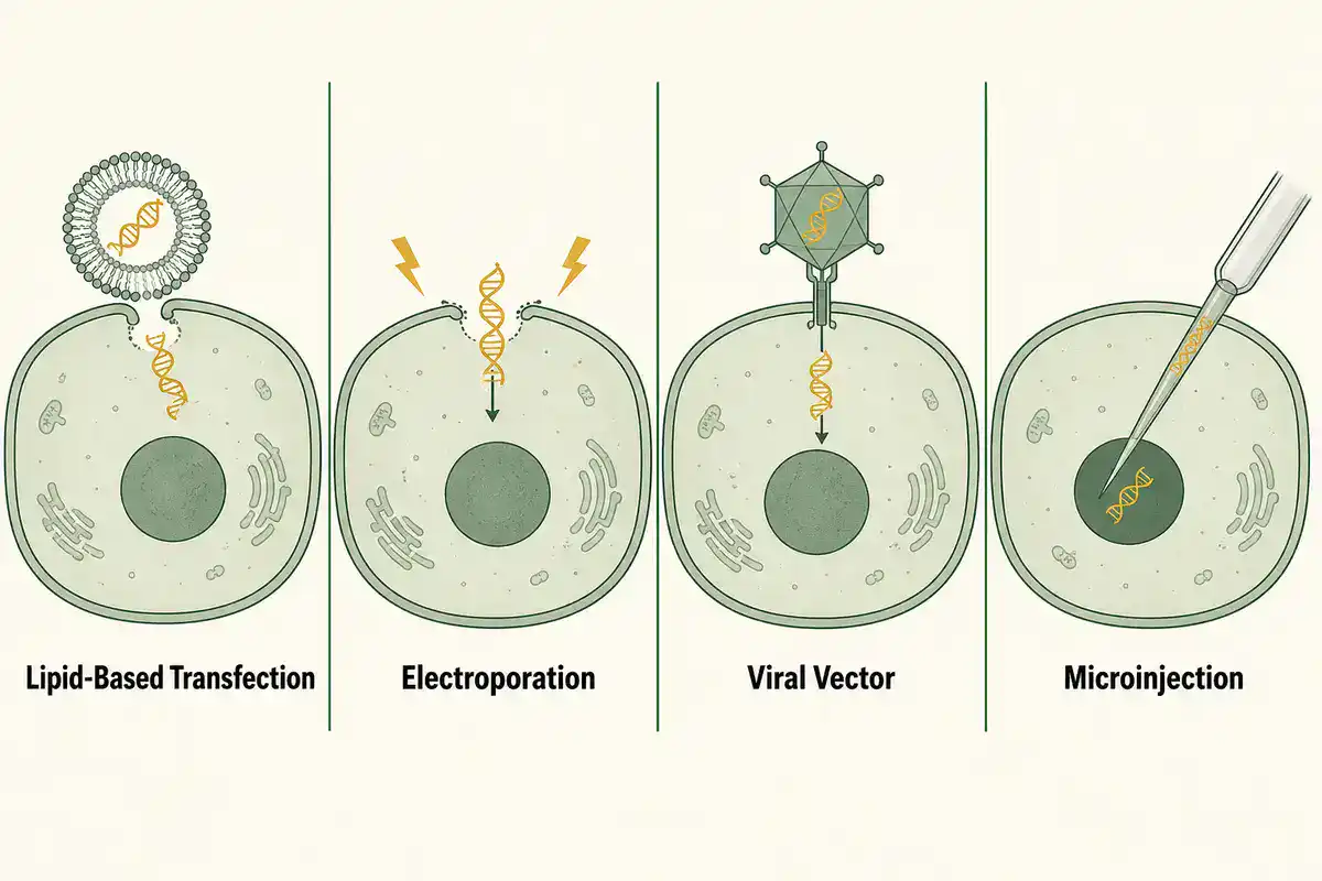
A useful transfection protocol should state the cell type, passage range, seeding density, confluence at transfection, nucleic acid amount, reagent ratio, medium conditions, incubation time, control groups, toxicity check, reporter or expression readout, and timing of analysis.
Transfection failure is often caused by the wrong cell density, poor cell health, toxic reagent conditions, low-quality plasmid or RNA, unsuitable medium, incorrect reagent ratio, or checking the result too early or too late. A good protocol should make these variables visible before the experiment starts.
Immunofluorescence and Cell Staining Protocols
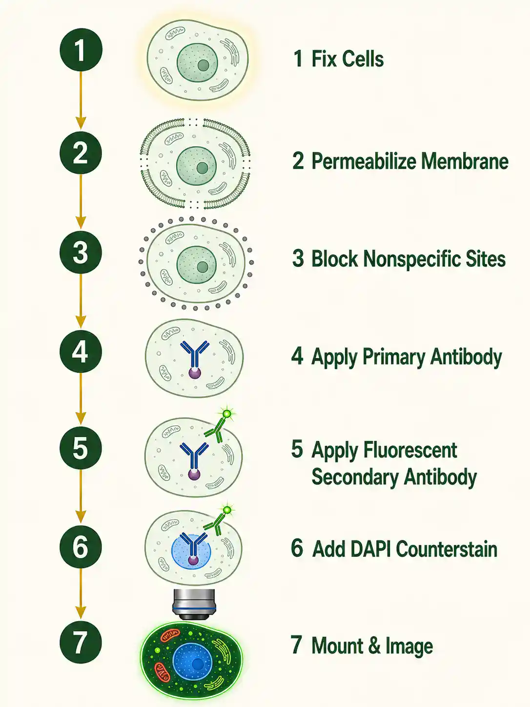
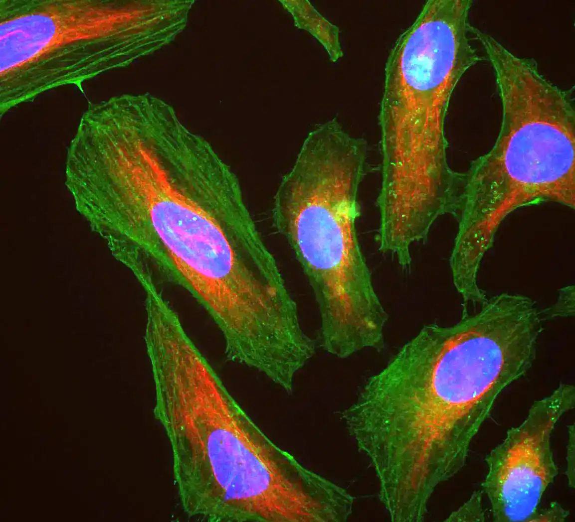
Immunofluorescence protocols use antibodies and fluorescent labels to detect proteins, structures, or cellular markers. They are widely used to study protein localization, organelles, cytoskeleton, cell identity, signaling, cell cycle, infection, and treatment response.
A useful immunofluorescence protocol should include fixation method, permeabilization conditions, blocking conditions, primary antibody, secondary antibody, nuclear stain if used, washing steps, mounting medium, imaging settings, controls, and analysis plan. Fixation and permeabilization are not neutral choices. They can preserve some structures while damaging or masking others.
Common controls include no-primary-antibody control, secondary-only control, known positive sample, known negative sample, isotype control when appropriate, and marker colocalization controls. Without controls, a bright image can still be misleading.
For microscopy-based cell biology, image acquisition should be treated as part of the protocol. Exposure time, objective lens, laser intensity, detector gain, focus plane, scale bar, file format, and image processing rules should be documented before comparing samples.
Flow Cytometry and Cell Sorting Protocols
Flow cytometry protocols measure properties of individual cells in a flowing sample. Flow cytometry can analyze cell size, internal complexity, viability, fluorescent markers, cell cycle, apoptosis, immune cell identity, and reporter expression.
NCBI Bookshelf describes flow cytometry as a technique that measures physical and chemical properties of cells by analyzing light scattering and fluorescence emission. A widely cited review explains that flow cytometers use lasers to generate scattered and fluorescent light signals that are read by detectors. See Flow Cytometry Blood Cell Identification and Flow Cytometry: An Overview.
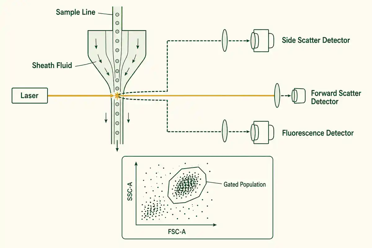
A strong flow cytometry protocol should include sample preparation, single-cell suspension quality, viability dye, antibody panel, staining buffer, compensation controls, fluorescence-minus-one controls when needed, gating strategy, instrument settings, event count, doublet exclusion, and data analysis plan.
Cell sorting is a specialized use of flow cytometry that physically separates selected cells. Sorting can enrich specific cell types or remove dead cells, but it also adds stress, biosafety concerns, instrument variables, and sample-quality requirements. For example, 10x Genomics notes that flow cytometry-based sorting can enrich specific cell types and remove dead cells for single-cell workflows. See Flow Cytometry Guidance for Single Cell Protocols.
Microscopy and Live-Cell Imaging Protocols
Microscopy protocols help researchers observe cells, organelles, proteins, cytoskeletal structures, membranes, and dynamic processes. Common approaches include brightfield microscopy, phase contrast, fluorescence microscopy, confocal microscopy, time-lapse imaging, and live-cell imaging.
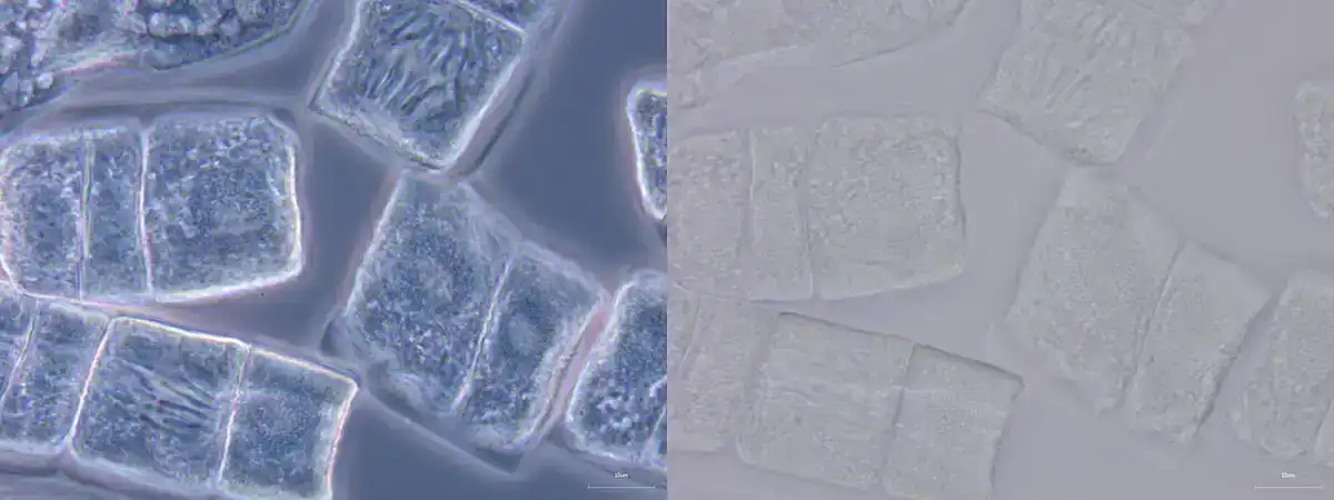
A good microscopy protocol should state sample preparation, plate or slide format, coating, fixation if used, stain or fluorescent marker, objective lens, magnification, exposure or laser settings, image format, scale bar, number of fields, number of cells, and analysis method.
Live-cell imaging requires extra care because cells must remain healthy during observation. Temperature, CO2, humidity, light exposure, phototoxicity, focus drift, medium evaporation, and imaging frequency can all affect the result.
A visually attractive image is not automatically a good data point. A useful imaging protocol should define how images will be collected, which cells or fields will be excluded, how signal will be measured, and whether image processing changes intensity, contrast, or interpretation.
Cell Cycle, Apoptosis and Proliferation Assays
Many cell biology protocols measure whether cells are dividing, arrested, dying, or responding to a treatment. These methods are common in cancer biology, toxicology, drug screening, developmental biology, immunology, and tissue engineering.
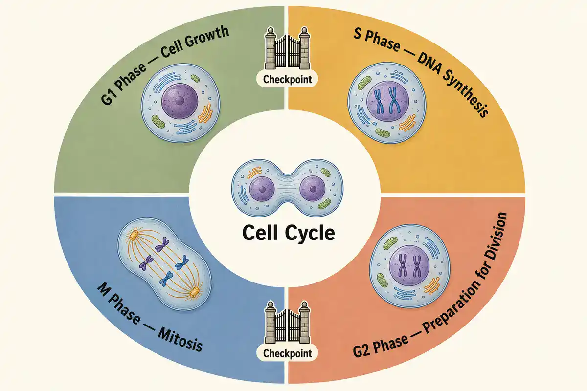
Cell cycle protocols may use DNA-binding dyes, EdU or BrdU incorporation, mitotic markers, flow cytometry, imaging, or reporter systems. They help estimate how cells are distributed across phases such as G1, S, G2, and M, depending on the assay design.
Apoptosis protocols may measure membrane changes, caspase activity, DNA fragmentation, mitochondrial membrane potential, Annexin V staining, or morphological changes. A single marker may not capture every form of cell death, so method choice matters.
Annexin V/PI Apoptosis Calculator
Proliferation assays can measure cell number, DNA synthesis, metabolic activity, confluence, colony formation, or label dilution. These readouts do not always mean the same thing. A treatment may lower metabolic signal without killing cells, or slow division without causing immediate cell death.
Migration, Invasion and Wound Healing Assays
Cell migration protocols measure how cells move. These methods are used in wound healing research, cancer biology, developmental biology, immunology, tissue repair, angiogenesis, and cell signaling studies.
Common approaches include scratch assays, transwell migration assays, invasion assays through matrix-like barriers, time-lapse imaging, and single-cell tracking. A good migration protocol should control for cell density, proliferation, serum conditions, matrix coating, scratch width if used, imaging interval, and measurement method.
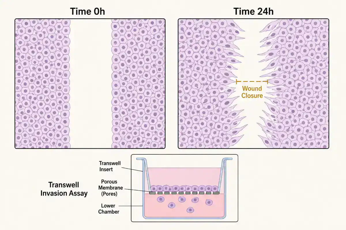
One common mistake is confusing migration with proliferation. If the assay does not control for cell division, faster wound closure may reflect increased growth rather than increased movement. Good protocols make that distinction clear.
How to Choose the Right Cell Biology Protocol
Do not choose a cell biology protocol only because it appears first in search results. Choose it because it matches your cell type, culture model, readout, instrument, treatment, safety requirements, and biological question.
Before using any protocol, check these points:
- Cell type: adherent, suspension, primary, immortalized, stem cell, immune cell, neuronal cell, epithelial cell, cancer cell, organoid, or co-culture.
- Cell state: passage number, confluence, viability, differentiation state, cell cycle state, contamination status, and recent treatment history.
- Goal: culture maintenance, imaging, protein localization, viability, transfection, flow cytometry, migration, apoptosis, proliferation, or cell sorting.
- Readout: microscopy image, fluorescence intensity, cell count, absorbance, luminescence, flow cytometry plot, qPCR result, western blot, or functional response.
- Controls: untreated control, vehicle control, positive control, negative control, staining control, viability control, no-primary control, compensation control, or loading control.
- Equipment: biosafety cabinet, incubator, microscope, centrifuge, cell counter, plate reader, flow cytometer, imaging system, or culture vessel format.
- Safety: cell source, infectious risk, recombinant material, viral vectors, human-derived material, chemical exposure, sharps, and waste disposal.
- Verification: know how success will be checked before beginning the protocol.
Common Mistakes in Cell Biology Protocols
Cell biology methods often fail for ordinary reasons. The assay may be valid, but the cells may not be healthy, consistent, or appropriate for the question.
- Using unhealthy cells: stressed, over-confluent, under-recovered, or contaminated cells can distort results.
- Ignoring passage number: cells can change behavior with repeated passaging or long culture time.
- Skipping contamination checks: mycoplasma and other contaminants can alter growth, metabolism, gene expression, and assay response.
- Changing seeding density casually: many cell assays depend strongly on starting cell number and confluence.
- Confusing viability with metabolism: some assays report metabolic activity rather than direct live-cell count.
- Using weak controls: without positive and negative controls, staining, transfection, viability, and flow results are hard to interpret.
- Overprocessing images: contrast, brightness, thresholding, and background subtraction can change interpretation if not applied consistently.
- Overlooking antibody specificity: immunofluorescence and flow cytometry depend on antibody performance and proper controls.
- Assuming one protocol fits every cell line: cell type, growth conditions, and assay sensitivity can change the workflow.
- Not documenting deviations: small changes in timing, media, temperature, passage number, or cell density can explain major result differences.
Cell Biology Calculators and Lab Tools
Cell biology protocols often depend on calculations. A wrong cell count, viability estimate, dilution, seeding density, reagent concentration, or transfection ratio can affect the experiment before the assay begins.
Useful cell biology calculators include:
- cell counting calculator
- hemocytometer calculator
- cell viability calculator
- cell seeding density calculator
- cell dilution calculator
- passage ratio calculator
- population doubling time calculator
- transfection reagent calculator
- multiplicity of infection calculator
- flow cytometry percentage calculator
- scratch assay migration calculator
- fluorescence intensity normalization calculator
- cell proliferation index calculator
- plate layout calculator
- serial dilution calculator
BioExplorer's Biology Tools and Calculators hub is building free browser-based tools by branch of biology. The existing Genetics and Inheritance Tools section includes inheritance and population genetics tools, while the Botany Tools section supports plant science workflows. As BioExplorer expands its cell biology tools, this page can link directly to cell counting, seeding density, viability, transfection, and assay-planning calculators.
Trusted Cell Biology Protocol Resources
Use BioExplorer as a guide, but always check your institution's approved SOPs, biosafety rules, manufacturer instructions, and supervisor guidance before performing real laboratory work. These external resources are useful starting points for cell biology protocols and method background:
- Nature Protocols: peer-reviewed protocol articles with design advice, troubleshooting, data analysis, limitations, and result interpretation.
- Bio-protocol: peer-reviewed open access protocol journal focused on improving research reproducibility.
- protocols.io: a platform for creating, versioning, sharing, and running reproducible research methods.
- ATCC Animal Cell Culture Guide: guidance on media, subculturing, cryopreservation, and contamination in animal cell culture.
- Gibco Cell Culture Basics Handbook: practical background on aseptic technique, contamination control, cell culture supplies, and routine culture handling.
- A Beginner’s Guide to Cell Culture: practical advice on cell culture quality, contamination, mycoplasma, and workflow issues.
- NCBI Bookshelf: Flow Cytometry Blood Cell Identification: overview of flow cytometry as a method for measuring light scattering and fluorescence properties of cells.
- Flow Cytometry: An Overview: review of flow cytometry principles, lasers, scattered light, fluorescence signals, and detectors.
- 10x Genomics Flow Cytometry Guidance: guidance on using flow cytometry and cell sorting to enrich cell types and remove dead cells for single-cell workflows.
- CDC Biosafety in Microbiological and Biomedical Laboratories: biosafety guidance based on protocol-driven risk assessment in biomedical and microbiological laboratories.
Safety and Responsibility
Cell biology protocols can involve living cells, human-derived material, animal-derived material, primary tissue, recombinant DNA, viral vectors, chemical stains, fixatives, disinfectants, sharps, centrifuges, UV light, biosafety cabinets, incubators, and specialized instruments. This page is educational. It does not replace formal training, institutional SOPs, biosafety approval, chemical safety guidance, or supervision by qualified personnel.
The CDC's biosafety guidance emphasizes protocol-driven risk assessment because no single document can identify every possible combination of risks and mitigation steps in biomedical laboratories. For any protocol involving infectious agents, human material, viral vectors, recombinant organisms, animal material, hazardous chemicals, or regulated samples, use approved institutional procedures and safety oversight.
Related BioExplorer Resources
- Biology Methods and Protocols: main hub for biology lab methods, protocol categories, safety notes, and trusted resources.
- Cell Biology: guide to cells, organelles, cell signaling, cell division, and cell-based experiments.
- Cell Biology Glossary: clear definitions of cells, organelles, mitosis, meiosis, membranes, cytoplasm, cytoskeleton, cell cycle, and related terms.
- Molecular Biology: DNA, RNA, proteins, gene expression, molecular mechanisms, and related research methods.
- Molecular Biology Methods and Protocols: protocols for DNA, RNA, PCR, cloning, gel electrophoresis, sequencing prep, and molecular analysis.
- Biochemistry Methods and Protocols: methods for protein assays, SDS-PAGE, western blotting, enzyme kinetics, chromatography, and protein purification.
- Biology Tools and Calculators: free browser-based biology calculators and educational tools.
Differences Between Mitosis and Meiosis
Frequently Asked Questions
Cell biology protocols are written laboratory workflows for growing, maintaining, counting, staining, imaging, treating, and analyzing cells under defined conditions.
Common cell biology protocols include cell culture, aseptic technique, passaging, cryopreservation, cell counting, viability assays, transfection, immunofluorescence, microscopy, flow cytometry, apoptosis assays, cell cycle analysis, and migration assays.
Aseptic technique reduces the chance that bacteria, fungi, mycoplasma, or other contaminants enter a cell culture. Contamination can change cell behavior, damage experiments, and make results unreliable.
Cell counting estimates how many cells are present. Cell viability estimates how many cells are alive or functionally active according to the assay used. A sample can have many cells but low viability, or fewer cells with high viability.
Useful immunofluorescence controls can include no-primary-antibody control, secondary-only control, known positive sample, known negative sample, isotype control when appropriate, and consistent imaging settings across samples.
Flow cytometry measures properties of single cells in a flowing sample, including light scatter and fluorescence. It can be used to study cell populations, marker expression, viability, cell cycle, apoptosis, and sorting of selected cells.
Not always. Online protocols vary in quality and may not match your cell type, biosafety level, equipment, reagents, or institution's rules. For real lab work, follow approved SOPs, safety guidance, manufacturer instructions, and supervisor instruction.
Cite this page
BioExplorer. (2026, August 26). Cell Biology Protocols. https://www.bioexplorer.net/methods_and_protocols/cell_biology/