Pathology: The Study of Disease
Pathology is the branch of biology and medicine that studies disease. It examines what goes wrong in cells, tissues, organs, blood, body fluids, genes, and immune responses when normal function is disrupted.
Pathology is where disease leaves evidence. A swollen lymph node, abnormal blood count, biopsy sample, tumor margin, infected tissue, damaged kidney, inflamed lung, genetic mutation, or autopsy finding can all become clues. The pathologist’s work is to turn those clues into a diagnosis, explanation, or research insight.
Pathology connects closely with anatomy, cell biology, molecular biology, biochemistry, physiology, immunology, microbiology, genetics, pharmacology, and parasitology.
Pathology Guide:
- Disease Leaves a Trail
- Pathology Definition and Meaning
- The Evidence Pathologists Read
- The Four Questions Behind Disease
- Anatomic Pathology and Clinical Pathology
- What Happens to a Biopsy?
- Cancer Pathology: Naming the Enemy Precisely
- Inflammation, Infection, and Tissue Injury
- The Laboratory Side of Pathology
- Molecular Pathology and Precision Medicine
- Pathology Is Also Research
- History of Pathology: Turning Points That Changed Diagnosis
- Tools Pathologists Use
- Pathology Careers
- Related BioExplorer Resources
- Recommended Pathology Resources
- Pathology FAQs
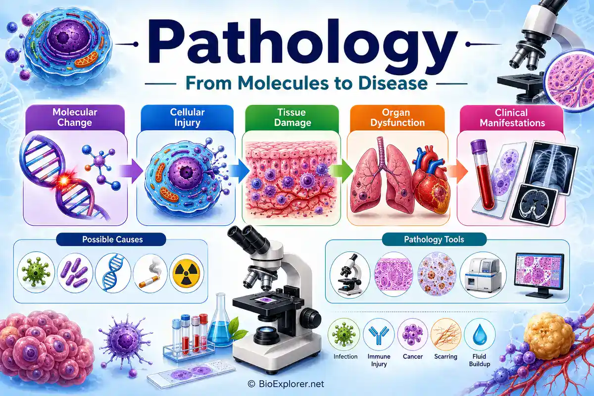
Disease Leaves a Trail
A disease rarely appears as one isolated fact. It leaves a trail across different levels of biology. A mutation can change a protein. A damaged protein can alter a cell. Altered cells can injure tissue. Damaged tissue can change organ function. That change may show up as symptoms, lab results, imaging findings, or microscopic patterns.
This is the heart of pathology. It connects the visible problem with the hidden mechanism. A cough may be linked to inflamed airways, infection, immune injury, cancer, fluid buildup, or tissue scarring. A high white blood cell count may point toward infection, inflammation, leukemia, stress, medication effects, or bone marrow disease. Pathology helps sort evidence from noise.
For biology students, pathology is useful because it shows normal biology under stress. To understand disease, you need normal cells, normal tissues, normal genes, normal immune defense, and normal physiology first.
Pathology Definition and Meaning
A practical pathology definition is: the scientific study of disease, including its causes, mechanisms, structural changes, functional effects, diagnosis, and consequences.
Pathology can refer to a medical specialty, a laboratory discipline, or a wider biological study of disease processes. In medicine, pathologists help diagnose disease by examining tissues, cells, blood, body fluids, molecular tests, microorganisms, and sometimes whole organs or bodies after death.
In biology research, pathology is also about pathogenesis, which means how disease develops. A pathologist or disease researcher may ask what caused the injury, what cells responded, what molecules changed, what tissue pattern appeared, and what that means for the organism.
The Evidence Pathologists Read
Pathology is not based on one kind of sample. Different diseases leave different evidence, so pathology uses different materials and methods.
| Evidence Type | What It Can Show | Example Use |
|---|---|---|
| Biopsy Tissue | Cell arrangement, tissue architecture, inflammation, infection, scarring, or cancer. | Diagnosing a breast lump, colon polyp, skin lesion, or liver disease. |
| Surgical Specimen | Disease extent, margins, organ involvement, and tumor features. | Checking whether a tumor was fully removed. |
| Blood Sample | Cell counts, abnormal cells, clotting changes, chemistry, infection markers, and immune findings. | Investigating anemia, leukemia, infection, bleeding, or organ damage. |
| Body Fluid | Cells, organisms, proteins, crystals, or chemical changes in fluid. | Testing cerebrospinal fluid, pleural fluid, urine, joint fluid, or abdominal fluid. |
| Cytology Sample | Individual cells or small cell groups. | Pap tests, thyroid needle aspiration, urine cytology, or fluid cytology. |
| Microbiology Sample | Bacteria, fungi, parasites, or viruses. | Identifying infection from blood, sputum, tissue, stool, or wound samples. |
| Molecular Test | DNA, RNA, gene variants, rearrangements, or pathogen genetic material. | Cancer classification, inherited disease testing, or infection detection. |
| Autopsy Findings | Whole-body disease patterns and cause of death. | Understanding unexpected death, disease complications, or treatment effects. |
The Four Questions Behind Disease
Pathology often organizes disease around four basic questions. This makes it different from simply naming a condition.
- Cause: What started the disease? Examples include infection, mutation, toxin, injury, immune reaction, poor blood supply, or inherited defect.
- Mechanism: How does the disease develop? This is the pathogenesis.
- Structural change: What changed in cells, tissues, organs, or body fluids?
- Functional result: How do those changes affect symptoms, lab tests, organ function, treatment, or outcome?
A diagnosis becomes more useful when these layers are connected. For example, a bacterial pneumonia diagnosis is stronger when the pathologist can connect infection, inflammation, lung tissue changes, organism detection, and clinical severity.
Top 14 Most Infectious and Deadliest Diseases Caused By Bacteria
Anatomic Pathology and Clinical Pathology
Modern pathology is often divided into anatomic pathology and clinical pathology. They overlap, but they read different kinds of evidence.
| Area | Main Focus | Common Examples |
|---|---|---|
| Anatomic Pathology | Diagnosis from organs, tissues, cells, and whole-body examination. | Biopsies, surgical specimens, autopsies, cytology, tumor margins, histology. |
| Clinical Pathology | Diagnosis from blood, urine, body fluids, chemistry, microbiology, hematology, and lab testing. | Blood counts, cultures, chemistry panels, coagulation tests, transfusion testing, molecular tests. |
| Histopathology | Microscopic study of diseased tissue architecture. | Cancer diagnosis, inflammation patterns, organ injury, fibrosis, infection. |
| Cytopathology | Study of individual cells or small groups of cells. | Pap tests, fine-needle aspiration, fluid cytology, thyroid cytology. |
| Molecular Pathology | Diagnosis using DNA, RNA, proteins, mutations, and molecular markers. | Cancer mutations, inherited disease testing, pathogen detection, targeted therapy markers. |
| Forensic Pathology | Investigation of death, injury, and disease in legal contexts. | Autopsy, cause of death, injury pattern analysis, toxicology correlation. |
What Happens to a Biopsy?
A biopsy is one of the most familiar gateways into pathology. A small piece of tissue is removed from the body and sent to a pathology laboratory. What happens next is careful, stepwise, and highly controlled.
- Collection: A clinician removes tissue through a needle, endoscope, surgery, or another procedure.
- Fixation: The tissue is preserved to reduce breakdown and maintain structure.
- Gross examination: The pathologist or trained professional examines size, shape, color, margins, and visible abnormalities.
- Processing: The tissue is embedded, usually in paraffin, so thin sections can be cut.
- Sectioning: Very thin slices are placed on glass slides.
- Staining: Dyes such as hematoxylin and eosin help reveal nuclei, cytoplasm, tissue patterns, and abnormal cells.
- Microscopic diagnosis: The pathologist examines the slide and interprets the findings.
- Special testing: Immunohistochemistry, molecular tests, special stains, or other tests may be added when needed.
- Report: The pathology report communicates the diagnosis and key findings to the treating clinician.
This process is why pathology reports matter so much in cancer diagnosis, transplant care, inflammatory disease, infection, and many other medical decisions.
Cancer Pathology: Naming the Enemy Precisely
Cancer pathology is one of the clearest examples of why pathology matters. A tumor is not treated only by where it appears. Pathologists help determine what type of tumor it is, whether it is benign or malignant, how aggressive it looks, whether it has invaded nearby tissue, whether margins are clear, and which molecular features may guide treatment.
A cancer pathology report may include tumor type, grade, stage-related features, margin status, lymph node involvement, biomarkers, receptor status, mutation testing, and other details. These findings can influence surgery, chemotherapy, radiation therapy, immunotherapy, targeted therapy, follow-up, and prognosis.
Inflammation, Infection, and Tissue Injury
Not every pathology case is cancer. Many diseases involve inflammation, infection, degeneration, immune attack, metabolic injury, poor blood supply, scarring, or toxic damage. A pathologist may see neutrophils in acute infection, granulomas in certain chronic diseases, necrosis after severe injury, fibrosis after long-term damage, or immune deposits in autoimmune disease.
Pathology helps separate look-alike problems. Fever and swelling may come from infection, immune disease, drug reaction, tissue death, or cancer. Tissue patterns, stains, cultures, molecular tests, and clinical information help narrow the cause.

Is MRSA Contagious? Explore the Etiology of MRSA Infection
The Laboratory Side of Pathology
Clinical pathology turns body fluids and laboratory results into medical information. This includes blood counts, chemistry values, clotting tests, cultures, urinalysis, transfusion testing, flow cytometry, molecular diagnostics, and many other tests.
A laboratory value is not just a number. High potassium, low hemoglobin, abnormal liver enzymes, increased white blood cells, positive blood culture, or abnormal clotting time all need biological interpretation. Pathology links the result to the patient, specimen quality, disease process, and clinical question.
| Lab Area | What It Studies | Example |
|---|---|---|
| Hematology | Blood cells, bone marrow, clotting, and blood disorders. | Anemia, leukemia, platelet disorders, clotting abnormalities. |
| Clinical Chemistry | Chemical substances in blood and body fluids. | Kidney function, liver enzymes, glucose, electrolytes, hormones. |
| Microbiology | Microorganisms that cause infection. | Bacterial culture, fungal identification, antimicrobial susceptibility. |
| Transfusion Medicine | Blood groups, compatibility, blood products, and transfusion safety. | Crossmatching blood before transfusion. |
| Immunopathology | Immune patterns in disease. | Autoimmune markers, immune deposits, allergy-related testing. |
| Molecular Diagnostics | DNA, RNA, mutations, rearrangements, and pathogen genetic material. | Cancer mutations, inherited variants, viral load testing. |
| Clinical Informatics | Data systems, lab information, quality, and diagnostic workflows. | Managing results, reference ranges, decision support, and lab data quality. |
Molecular Pathology and Precision Medicine
Molecular pathology studies disease through DNA, RNA, proteins, mutations, gene rearrangements, expression patterns, and molecular markers. It is especially important in cancer, inherited disease, infectious disease, pharmacogenomics, and transplant medicine.
In cancer care, molecular pathology can help identify mutations that make a tumor more likely to respond to a targeted therapy. In infectious disease, molecular tests can detect pathogen genetic material. In inherited disease, genetic testing can help identify variants linked to disease risk or diagnosis.
This does not replace tissue pathology. It adds another layer. The most useful diagnosis often combines tissue pattern, cell type, clinical history, imaging, immunostains, and molecular results.
Pathology Is Also Research
Pathology is not only diagnostic. Investigative pathology studies disease mechanisms. Researchers may study why cells die, how inflammation damages organs, how tumors invade, why infections spread, how fibrosis forms, how blood vessels change, or how genetic variants alter tissue function.
Experimental pathology often works with animal models, cell cultures, organoids, tissue sections, imaging, molecular assays, and human specimens. The goal is to understand disease deeply enough to improve diagnosis, prevention, and treatment.
History of Pathology: Turning Points That Changed Diagnosis
The history of pathology changed when disease moved from vague symptoms to anatomical locations, then to cells, microbes, stains, blood groups, cytology, antibodies, DNA, and digital images. These are selected turning points, not a full timeline.
| Year | Discovery or Contribution | Why It Matters |
|---|---|---|
| 1761 | Giovanni Battista Morgagni published De Sedibus et Causis Morborum per Anatomen Indagatis. | Helped connect symptoms with organ-based lesions and strengthened pathological anatomy. |
| 1858 | Rudolf Virchow published Cellular Pathology. | Moved disease understanding toward cells and microscopic tissue change. |
| 1876 to 1882 | Robert Koch connected specific bacteria with specific infectious diseases, including anthrax and tuberculosis. | Helped establish bacteriology as a foundation for infectious disease pathology. |
| 1884 | Hans Christian Gram developed the Gram stain. | Made bacteria more visible in tissue and later became a key method for classifying bacteria. |
| 1900 to 1901 | Karl Landsteiner discovered the ABO blood groups. | Transformed transfusion medicine and laboratory compatibility testing. |
| 1941 | George Papanicolaou and Herbert Traut published work showing the diagnostic value of vaginal smears for uterine cancer. | Helped establish exfoliative cytology and the Pap test as a cancer-screening tool. |
| 1941 | Albert Coons and colleagues introduced fluorescent antibody labeling. | Opened the way for immunofluorescence and later antibody-based tissue diagnosis. |
| 1983 | Kary Mullis developed polymerase chain reaction, or PCR. | Made targeted DNA amplification practical and later transformed molecular diagnostics. |
| 2017 | The FDA permitted marketing of a whole-slide imaging system for primary diagnostic use in surgical pathology. | Marked an important regulatory milestone for digital pathology in clinical diagnosis. |
Tools Pathologists Use
Pathology tools are designed to make disease visible, measurable, and interpretable. Some tools show structure. Others show proteins, genes, microbes, chemistry, or cell behavior.
- Gross examination: Careful visual and physical examination of organs or tissues.
- Light microscopy: Study of stained tissue or cell samples using a microscope.
- Hematoxylin and eosin staining: A common tissue stain that highlights nuclei, cytoplasm, and tissue architecture.
- Special stains: Stains that highlight organisms, minerals, mucus, iron, fibrosis, or other tissue features.
- Immunohistochemistry: Antibody-based staining used to detect proteins in tissue.
- Flow cytometry: Analysis of cells using markers, light scatter, and fluorescence.
- Microbial culture: Growth of microorganisms to identify infections and test susceptibility.
- Molecular testing: DNA, RNA, or gene-based testing for diagnosis and classification.
- Digital pathology: Scanning slides into digital images for viewing, consultation, measurement, education, or computational analysis.
Pathology Careers
Careers in pathology can follow the sample, the disease, the laboratory, or the research question. Some pathologists diagnose tissue. Others direct clinical laboratories, study blood diseases, investigate infections, perform autopsies, develop molecular tests, support cancer care, or study disease mechanisms.
- Anatomic pathologist: Diagnoses disease from tissues, organs, biopsies, surgical specimens, cytology, or autopsy material.
- Clinical pathologist: Directs and interprets laboratory testing of blood, urine, body fluids, chemistry, microbiology, and transfusion medicine.
- Histopathologist: Studies diseased tissue under the microscope.
- Cytopathologist: Diagnoses disease from individual cells or small cell groups.
- Molecular pathologist: Uses molecular tests to diagnose and classify disease.
- Hematopathologist: Studies blood, bone marrow, lymph nodes, leukemia, lymphoma, and related disorders.
- Forensic pathologist: Investigates cause and manner of death in legal and medical contexts.
- Veterinary pathologist: Studies disease in animals and supports veterinary medicine, wildlife health, and research.
- Experimental pathologist: Studies disease mechanisms in research settings.
- Pathology informatics specialist: Works with laboratory data, digital pathology, diagnostic systems, and computational tools.

How To Become A Pathologist?
Related BioExplorer Resources
Use these BioExplorer pages to connect pathology with disease biology, cells, tissues, infection, immunity, and molecular mechanisms:
- Anatomy
- Cell Biology
- Molecular Biology
- Biochemistry
- Physiology
- Immunology
- Microbiology
- Virology
- Parasitology
- Genetics
- Pharmacology
- Cellular Organization
- Building Blocks of Proteins
- Building Blocks of Nucleic Acids
Recommended Pathology Resources
These external resources are useful for learning about pathology, disease mechanisms, pathology reports, histopathology, molecular pathology, laboratory medicine, and pathology careers.
- Royal College of Pathologists: What Is Pathology? A clear introduction to pathology as the study of disease and its role in healthcare.
- Britannica: Pathology A concise reference on pathology, disease causes, structural changes, and history.
- College of American Pathologists A major professional organization for pathologists, laboratory quality, and pathology practice.
- American Society for Investigative Pathology A professional society focused on mechanisms of disease and experimental pathology.
- American Board of Pathology A certification resource for pathology training, primary certificates, and subspecialties.
- United States and Canadian Academy of Pathology A professional education and meeting resource for anatomic pathology.
- Pathology Outlines A widely used pathology reference for diseases, organs, microscopic features, and diagnostic terminology.
- University of Utah WebPath A long-standing educational pathology resource with images, tutorials, and self-study materials.
- National Cancer Institute: Pathology Definition A cancer-focused definition of pathology and its diagnostic role.
- SEER Training: Pathology A useful training resource for cancer pathology terminology and reports.
- FDA: First Whole-Slide Imaging System for Digital Pathology A regulatory milestone for digital pathology in primary diagnosis.
Pathology FAQs
Pathology is the scientific study of disease. It examines causes, mechanisms, cell and tissue changes, lab findings, diagnosis, and the effects of disease on the body.
A pathologist helps diagnose disease by examining tissues, cells, blood, body fluids, microorganisms, molecular tests, and sometimes autopsy findings. Pathologists also support research and laboratory medicine.
Pathology studies disease causes, tissue changes, lab findings, and diagnosis. Pathophysiology focuses more on how disease disrupts normal body function.
Anatomic pathology diagnoses disease by examining organs, tissues, biopsies, surgical specimens, cytology samples, and autopsy findings.
Clinical pathology diagnoses disease through laboratory testing of blood, urine, body fluids, chemistry, microbiology, hematology, transfusion medicine, and molecular tests.
Histopathology is the microscopic study of diseased tissue. It is commonly used to diagnose cancer, inflammation, infection, fibrosis, and organ injury.
Pathology is important because it explains what disease is, what caused it, how it affects cells and tissues, and how test results can guide diagnosis, treatment, research, and prevention.
Pathology careers include anatomic pathologist, clinical pathologist, histopathologist, cytopathologist, molecular pathologist, hematopathologist, forensic pathologist, veterinary pathologist, experimental pathologist, and pathology informatics specialist.
Cite this page
Bio Explorer. (2026, June 27). Pathology: The Study of Disease. https://www.bioexplorer.net/divisions_of_biology/pathology/