Cell Biology News of 2020: To have a healthy body, we need to have cells that work as intended. Starting from our earliest development, multiple cells work in concert, changing and dividing, creating complex organisms, and performing various functions.
The year 2020 was the time of an unprecedented health crisis. Understanding the inner body workings has become even more crucial than ever. There were multiple exciting discoveries made in 2020, including new information about stem cells, cell division, and even such a seemingly simple process as tasting flavors.
Top Cell Biology News of 2020
Here were present a collection of new insights about the tiny workers in our bodies.
1. A new spindle weaver found: researchers have uncovered novel facts about cell division mechanism – [USA, January 2020].

The division of the cell is one of the most crucial processes in its life cycle. The division depends on structures called microtubules – special rods made of a protein called tubulin.
Each microtubule is formed from a “seed” made of tubulin proteins coming together. This process is called microtubule nucleation. Spontaneous nucleation is too slow for the cell, so some proteins assist the creation of microtubules in making their formation more efficient.
One of such proteins is the γ-tubulin ring complexor γ-TURC.
A research team at the Princeton University has decided to recreate in the laboratory how the microtubules form branches during cell division. To do this, the scientists used an extract of the egg of the African clawed frog, Xenopus laevis .
Then they tried to record the events in real-time. Their experiment has shown that:
- Besides γ-TURC, two other proteins were essential for developing the microtubule network, augmin, and TRX2.
- TPX2 was the protein that played a central role in this process.
- TPX2 attaches itself to the starting microtubule and attracts both augmin and γ-TURC to this starting point.
- Without a concerted effort from augmin and TRX2, the protein γ-TURCcannot help form new microtubule branches.
- The role of TPX2 is of special interest as this protein is elevated in prostate cancer and several other cancers.
- This experiment is vital as the early events of microtubule formation were recreated for the first time.
This experiment is basically the proof of principle. A similar approach can be used to study cell division and form other types of microtubules.
- Reference: “Biochemical reconstitution of branching microtubule nucleation | eLife”. Accessed May 23, 2021. Link.
![]()
2. A sorter coming out of the nucleolus: A chromosome sorter protein complex discovered in the nucleolus – [Japan, July 2020].
The nucleus does not only contain chromosomes. There is a smaller structure called the nucleolus. It is basically composed of multiple droplet-like structures containing a mix of RNA, nucleotides, and proteins. When the cell functions normally, the nucleolus’ primary role is to produce ribosomes-specialized structures crucial for protein production.
Nucleolus can fall apart in case of stress or when the cell is preparing to divide. When nucleolus disassembles, it releases multiple proteins that can have diverse functions. These nucleolar proteins are now being actively studied.
Previously, it was discovered that one of the proteins present in the nucleolus, called NOL11, is crucial for normal cell division. If this protein is absent from the cell, the cell does not divide properly.
A team of Japanese scientists has carried out a series of experiments trying to understand the exact role of this previously understudied molecule.
- It was discovered that NOL11 binds to two other proteins called WD-protein 43 (WD-43) and Cirhin, forming a complex.
- This complex was called NWC.
- NWC forms during mitosis or cell division.
- When the scientists switched off the genes responsible for the proteins forming NWC, they have discovered that the chromosomes are not sorted equally during division.
- NWC also influences another essential protein that is active during cell division called Aurora B.
It is crucial to understand how disturbances to cell division occur. Multiple disorders are caused by the uneven sorting of chromosomes into the cell, including the well-known Down’s syndrome. Understanding what players participate in this complex process may be possible to prevent the creation of cells with the wrong chromosome number in the future.
Suggested Reading:
Can Animals Have Down Syndrome?
- Reference: “Identification of a novel nucleolar protein complex required for mitotic chromosome segregation through centromeric accumulation of Aurora B | nucleic acids Research | Oxford Academic”. Accessed May 23, 2021. Link.
![]()
3. Depolarizing for longer life: the ability to live longer in some species was linked to the “mild depolarization” mechanism of the mitochondria – [Germany-Russia-Switzerland, March 2020].
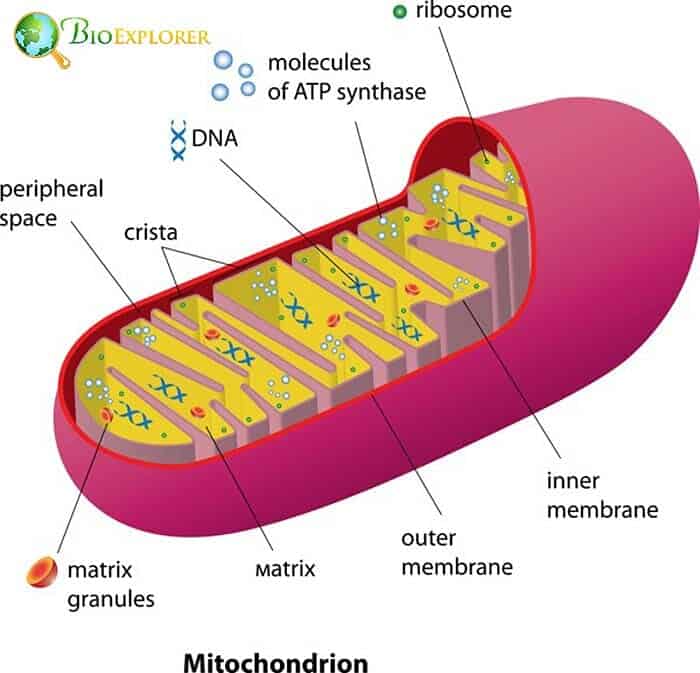
Cells cannot live without mitochondria. Mitochondria are molecular factories that produce energy currency – ATP. Like lumps of coal, ATP molecules are used to power up all the reactions in the cell.
Unfortunately, ATP production has side effects. Active mitochondria also leak substances that can quickly react with their surroundings called reactive oxygen species (ROS). ROS molecules in high quantities are destructive and contribute to slow decline and aging in the organism.
Recently, it was discovered that there is a mechanism in the cell that prevents mitochondria from producing ROS while continuing to produce ATP. Several enzymes called hexokinases, and creatine kinase can change the electric potential in the mitochondrial membrane. This leads to the decreased activity of mitochondria. This process is called mild depolarization.
Such a mechanism exists in many animals‘ cells. An international collaboration between German, Russian, and Swiss scientists has shown exciting results when the researchers have studied the activity of the mitochondria in the cells of mice, naked mole rats, and bats:
- In mice, the “mild depolarization” mechanism was active for several months in most organs.
- When mice grow to be 2.5 years old, the ability to depolarize mitochondria disappears from multiple cell types, including cells of the muscle, kidney, spleen, and brain.
- In naked mole rats and bats, the enzymes depolarizing mitochondria are active at low levels throughout their whole lives.
- Both naked mole rats and bats have longer lifespans compared to the laboratory mouse.
The scientists proposed that the mild depolarization mechanism is one of the factors that slows down aging in mammals. This discovery shows that the mechanisms preventing premature aging are complex. More research on the control of mitochondria activity is needed.
- Reference: “Mild depolarization of the inner mitochondrial membrane is a crucial component of an anti-aging program | PNAS”. Accessed May 23, 2021. Link.
![]()
4. Passing the force around – the scientists found how muscle cells send information about mechanical impacts – [Germany, December 2020].
The information about mechanical forces is passed down in cells through a complex mechanism. One of the proteins involved in passing around this information is called vinculin.
Recently, it was discovered that vinculin has a variant – a protein slightly different in a structure called metavinculin.
This protein also organizes other structures involved in spreading mechanical information differently. The function and properties of this variant were not well studied because there was no method to measure the mechanical forces inside the cell.
- A team of scientists at the University of Münster has developed new sensors to detect forces that can measure up to 10-12 newtons or piconewtons.
- It was found that metavinculin spreads mechanical forces less efficiently than vinculin.
- Metavinculin acts as a buffer, helping muscle cells withstand a sudden strong impact.
- The researchers have developed a mouse model that lacks the metavinculin genes alone.
- The modified mice did not have a higher risk of heart disorder.
- Using this model, the researchers have also disproven the assumption that the absence of metavinculin could contribute to the development of heart muscle hypertrophy.
The new approach and model developed by the team can help understand how muscle cells work in greater detail in the future.
- Reference: “Metavinculin modulates force bacterial transduction in cell adhesion sites | Nature Communications”. Accessed May 23, 2021. Link.
![]()
5. How to make a nucleus bigger: A protein helping cell nuclei grow found – [Germany, September 2020].
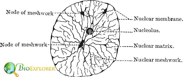
When cells divide, the containers for genetic material -the cell nuclei – get dissolved. The DNA gets condensed into the tightly packed chromosomes. After two new daughter cells form, each with their own chromosomes, a new nucleus would be forming in each of the new cells.
The new nucleus is initially relatively small. That could pose a problem, as the DNA previously packed into chromosomes needs to be partially unwound so the cell could pick up “orders” from the genes.
This means that nuclei need to grow to accommodate all the loops of DNA and accompanying proteins that work there.
Previously, it was not clear what happens inside the nucleus right after the cell division. The research team from the University of Freiburg has obtained fractions of cells that have just finished cell division. They have found that:
- A protein called ACTN4 has appeared in the newly formed nucleus.
- ACTN4 interacted with other proteins in the nucleus, particularly actin that plays a vital role as a support protein and contributes to growth.
- ACTN4 interacts with actin, helping form actin bundles that can act as an inner support framework.
- In cells where the ACTN4 gene was not functioning correctly, the nuclei could not grow while the cells themselves were of the regular size.
The researchers hope to study these processes closer to understand how those protein interactions make the cell nuclei grow.
- Reference: “Postmitotic expansion of cell nuclei requires nuclear actin filament bundling by α‐actinin 4 | EMBO reports”. Accessed May 23, 2021. Link.
![]()
6. Deathly traffic jams: Ribosome collisions can lead to the suicide of the cell – [USA, June 2020].
Ribosomes are crucial components of the cell. They serve as assembly lines, where proteins are made based on the blueprints provided by the mRNA. Sometimes, when the initial “blueprint” is faulty or some other issue, ribosomes can get stuck together.
Such traffic jams are dangerous – the protein production stalls, as does the transport within the cell.
Researchers from the John Hopkins University, Baltimore, MD, have focused on the mechanisms that the cell uses to protect itself from the consequences of such “traffic problems“.
- The researchers have subjected the test cells to various factors that block the normal function of ribosomes and make them stick together.
- When ribosomes collide, a protein called ZAK attaches itself to ribosomes coupled together.
- A phosphate group is attached to the ZAK protein, which changes its shape and activates the protein.
- If the numbers of collided ribosomes are low, another protein called GGN2 is attached to the complex with ribosomes and ZAK.
- Attachment of the GGN2 protein initiates a series of proteins participating in the integrated stress response – a cellular form of damage control.
- If there are many stuck ribosomes, a chain of events is activated that eventually leads to cell suicide.
- This process can be potentially regulated with drugs. So it could be possible to treat patients to kill dangerous cells (for example, in cancer) or protect the body’s cells from dying.
- Reference: “Ribosome Collisions Trigger General Stress Responses to Regulate Cell Fate: Cell”. Accessed May 23, 2021. Link.
![]()
7. Exiling for survival: cells can kick out mitochondria to survive in harmful conditions. – [Japan, June 2020]
Animal cells depend on mitochondria. Without the energy these organelles produce, the cells cannot survive. Damaged mitochondria can also be dangerous for the cell, as they start leaking toxic substances – just like a faulty battery.
Previously, it was thought that cells just digest the faulty mitochondria to protect the whole cell. This process is called mitophagy.
Japanese scientists from Osaka University have described a completely different way of saving the cell from faulty mitochondria:
- The researchers have labeled the mitochondria with fluorescent markers.
- The labeled cells were exposed to an agent that damages mitochondria.
- Multiple photoshots of the cells were made at regular intervals to document the processes inside them.
- It was discovered that the cells push the damaged mitochondria outside.
- Sometimes the mitochondria are enclosed in bubbles; sometimes, they are just outed whole.
- It was also established that a gene called Parkin which blocks the process of mitochondrial exile.
- If the Parkin is damaged, more mitochondria get pushed out of the cell than normal.
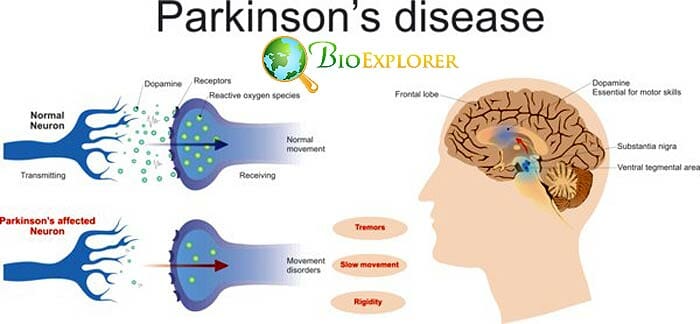
- Mutations in the Parkin gene are common in Parkinson’s disease.
- In patients with Parkinson’s disease, multiple mitochondria located outside the cells were found in cerebrospinal fluid.
This extracellular exclusion of mitochondria is a widespread mechanism of quality control of the normal mitochondria function. It is also of interest for medical research as this process may contribute to neurodegenerative diseases.
- Reference: “Full article: Alternative mitochondrial quality control mediated by extracellular release”. Accessed May 23, 2021. Link.
![]()
8. Superpowered stem cell discovered: A new type of stem cells found in early-stage embryos – [UK, August 2020].
One of the most fascinating aspects of embryonic development is that multiple organs arise from a limited number of cells. The embryo contains cells that are called stem cells.
Their main feature is the ability to divide and transform into multiple types of specialized cells. There are different types of stem cells. Among others, there are so-called) hematopoietic stem cells (HSC) that give rise to multiple cell types found in the bloodstream.
These HSCs can develop when the embryo is at its earliest stages. There is a unique location for them called the aorta-gonad mesonephros region. This region is located in the lower area of the tiny embryo, where the urogenital system would develop in the future.
Researchers from the University of Edinburgh, United Kingdom, specifically focused on early HSCs because they can play a crucial role in blood cancer treatment.
To study them better, the scientists transplanted embryonic cells into adult mice with specifically modified genotypes. They have found the following:
- First, hematopoietic stem cells appear in the embryo in the AGM region at the 32-41 days of development.
- Hematopoietic stem cells taken from embryos at different development stages can generate different quantities of cells.
- The highest number of new cells was generated from HSCs cells taken from the AGM region of 41-day embryos.
- Cells taken from this area at this point in time can produce up to 1, 600 daughter cells.
- This number is significantly lower for the stem cells found in umbilical cord blood.
The team intends to study the newly discovered type of cells, hoping to recreate it. With highly potent blood stem cells, treatment for blood cancers can become more efficient.
- Reference: “Vast Self-Renewal Potential of Human AGM Region HSCs Dramatically Declines in the Umbilical Cord Blood”. Accessed May 23, 2021. Link.
![]()
9. Peroxisome hidden secrets revealed with the help of plant cells: Unexpected hidden compartments discovered in an essential cellular structure – [USA, December 2020].
Sometimes cells need to destroy large molecules to get nutrients from them. Often hydrogen peroxide and other destructive substances are used in such reactions.
Therefore, the molecules to be destroyed and the substances used to digest the said molecules are hidden inside specialized bubbles called peroxisomes.
Previously, it was thought that peroxisomes are simple structures formed by a uniform, one-layer membrane. A recent study has shown it is not always so:
- A team of biochemists at Rice University, USA, studied peroxisomes in a model plant, Arabidopsis thaliana.
- To visualize the inner content of the peroxisomes, the researchers fused membrane proteins that usually can be found inside peroxisome membranes with bright reporter fluorescent proteins.
- When the fused proteins are built into new peroxisome, they can become visible, emitting fluorescent signals.
- It was discovered that membrane proteins were seen both in the outer membrane of the peroxisome and inside the peroxisome itself.
- It was particularly noticeable in large peroxisomes that are common in young plant seedlings.
- Additional inner membranes are formed in the large peroxisomes to better digest certain types of fatty acids.
- There are also different proteins sorted into different divided areas in the peroxisomes.
- Division of peroxisome into smaller chambers or compartments is likely not only present in plant cells.
- It was possible to study plant peroxisomes in greater detail because they are significantly larger than peroxisomes in animal cells.
This discovery overturns the previous concepts about peroxisome structure and function.
![]()
10. Tasting it all: New type of taste cells discovered in mice – [USA, August 2020].

Tasting various flavors is a complicated process. Our tongues contain different types of cells that work with taste:
- Support cells;
- Cells that detect bitter taste, sweet taste, and a type of flavor called umami.
- Sour and salty flavors are detected by the third type of cells.
Several years ago, researchers from the Center for Ingestive Behavior Research at the University of Buffalo, NY, USA, have found that two types of cells respond to bitter taste in mice.
In 2020, the team had decided to study these cells further in genetically modified mice and have discovered that:
- The new type of cells can respond to 4 tastes – bitter, sweet, sour, and umami.
- These cells use a different mechanism called a PLCβ3 pathway to detect bitter, sweet, and umami tastes.
- The scientists also discovered that both normal taste cells and the “universal” cells are needed to properly detect taste in food.
This discovery shows that deciphering taste is much more complex than previously thought.
- Reference: “A subset of broadly responsive Type III taste cells contribute to the detection of bitter, sweet and umami stimuli”. Accessed May 23, 2021. genetics/article?id=10.1371/journal.pgen.1008925″ target=”_blank”>Link.
![]()
Suggested Reading:
Top 10 Cell Biology Discoveries in 2019
The year 2020 has brought us new scientific insights into how our cells work. It is fascinating that new ways of controlling and managing such organelles as mitochondria and ribosomes were discovered. It is now known that cells can rewire their chemistry to survive with damaged mitochondria.
The cells also have a complex mechanism that repairs ribosome collisions. These trends are up-and-coming, as mitochondrial diseases are a very pertinent health problem.
![]()


