
Genetics Methods and Protocols

Genetics protocols are practical methods used to study genes, DNA, chromosomes, inheritance, genetic variation, mutations, gene expression, genotypes, pedigrees, populations, and genetic disease. These methods help researchers extract DNA, amplify target sequences, separate DNA fragments, genotype individuals, sequence genes or genomes, analyze variants, study inheritance patterns, and connect genetic results back to a biological question.
This page is a guide to major genetics methods and protocols used in research labs, teaching labs, biotechnology, molecular biology, genomics, population genetics, clinical genetics, evolutionary biology, microbiology, agriculture, conservation biology, and biomedical science. It explains what each method is for, what a good protocol should include, where mistakes usually happen, and which trusted resources can help you go deeper.
A useful genetics protocol is more than a list of steps. It should explain the sample type, DNA or RNA target, extraction method, assay design, controls, quality checks, contamination safeguards, data readout, interpretation rules, reporting limits, safety issues, and troubleshooting. Nature Protocols describes strong protocols as including reagents, equipment, timing, procedures, design advice, limitations, troubleshooting, data analysis, and result interpretation.
What Are Genetics Protocols?
Genetics protocols are written workflows for studying inherited information. They may be used to isolate DNA, amplify a gene region, detect a mutation, compare alleles, build a pedigree, genotype a plant or animal, identify a pathogen strain, study a population, sequence a genome, or interpret a genetic variant.
The method is the scientific technique. The protocol is the practical workflow for using that technique. For example, PCR is a method for amplifying a selected DNA sequence. A PCR protocol describes the template, primers, polymerase, thermal cycling design, positive control, negative control, expected product size, and how the amplified DNA will be checked or analyzed.
Genetics Protocols Guide
Use this page as a map for common genetics lab methods and genetic analysis workflows.
- DNA sample collection and storage
- genomic DNA extraction and quality checks
- PCR and target amplification
- agarose gel electrophoresis and fragment analysis
- restriction digestion and RFLP analysis
- genotyping and marker analysis
- Sanger sequencing and next-generation sequencing
- variant calling, annotation, and interpretation
- gene expression and RT-qPCR methods
- linkage analysis, association studies, and GWAS concepts
- pedigree analysis and inheritance-pattern interpretation
- population genetics and allele-frequency analysis
- CRISPR and genome-editing validation
- quality control, reporting, ethics, and troubleshooting
Core Genetics Methods at a Glance
The table below gives a quick map of important genetics methods and what each one helps measure or interpret.
| Method | Main Purpose | Common Readout |
|---|---|---|
| DNA extraction | Isolate DNA from cells, tissues, blood, saliva, plants, microbes, or environmental samples | DNA concentration, purity ratio, integrity, downstream PCR or sequencing success |
| PCR | Amplify a selected DNA region | Expected band size, amplification success, presence or absence of target |
| Gel electrophoresis | Separate DNA fragments by size | Band pattern, fragment length estimate, PCR product check, restriction digest pattern |
| Genotyping | Determine which alleles or variants are present at selected loci | Genotype call, allele call, marker pattern, zygosity |
| Sanger sequencing | Read a targeted DNA sequence | Chromatogram, base calls, sequence alignment, variant confirmation |
| Next-generation sequencing | Sequence many DNA fragments in parallel | Reads, coverage, variants, gene panel result, exome, genome, or targeted assay output |
| RT-qPCR | Measure relative or absolute gene expression from RNA-derived cDNA | Ct or Cq values, fold change, expression level, reference-gene normalization |
| RFLP analysis | Detect DNA variation that changes restriction-enzyme cutting patterns | Fragment pattern after digestion and gel electrophoresis |
| Pedigree analysis | Study inheritance patterns in families | Autosomal dominant, autosomal recessive, X-linked, mitochondrial, or more complex patterns |
| Population genetics analysis | Study allele frequencies, genetic diversity, and evolutionary forces in populations | Allele frequency, genotype frequency, heterozygosity, Hardy-Weinberg comparison, F-statistics |
DNA Sample Collection and Storage
Genetics begins with the sample. A strong genetic result depends on the quality, identity, and handling of the sample before any PCR, sequencing, or genotyping method begins.
DNA may come from blood, saliva, buccal swabs, hair roots, tissue biopsies, embryos, cultured cells, bacteria, fungi, plant leaves, seeds, museum specimens, environmental samples, or forensic material. Each sample type has different risks for degradation, contamination, inhibitors, mixed DNA, low yield, or mislabeling.
A useful sample-collection protocol should state the sample source, collection method, label format, storage condition, chain of custody if relevant, consent or permit requirements, contamination controls, and sample rejection criteria. For human genetics, ethical handling is part of the protocol, not an afterthought.
For environmental, forensic, ancient, or low-biomass samples, contamination control becomes especially important because small amounts of stray DNA can affect interpretation. Extraction blanks, collection controls, and careful documentation help distinguish real biological signal from handling artifacts.
Genomic DNA Extraction and Quality Checks
DNA extraction protocols isolate DNA from biological material. A good protocol breaks cells open, protects DNA from degradation, removes proteins and other contaminants, and produces DNA that is suitable for the next method.
DNA extraction is not one universal method. Plant tissues may contain polysaccharides and phenolic compounds. Soil samples may contain PCR inhibitors. Blood contains proteins and heme-related substances. Microbial cells may need stronger lysis. Formalin-fixed tissue can produce fragmented DNA. The extraction method should match the sample and the downstream use.
A useful DNA extraction protocol should state the sample type, lysis method, DNA-binding or precipitation strategy, wash steps, elution conditions, expected yield, DNA purity check, integrity check, storage conditions, and downstream assay. It should also state whether an extraction blank or negative control is included.
DNA quality can be checked by absorbance ratio, fluorescence-based quantification, gel electrophoresis, fragment analysis, or downstream performance. A high concentration number alone does not prove that DNA is usable. Inhibitors, fragmentation, carryover ethanol, protein contamination, or mixed samples can still cause problems.
PCR and Target DNA Amplification
PCR protocols, short for polymerase chain reaction protocols, amplify a selected DNA region. PCR is used in genotyping, mutation detection, cloning, microbial identification, forensic genetics, medical genetics, evolutionary studies, teaching labs, and sequencing preparation.
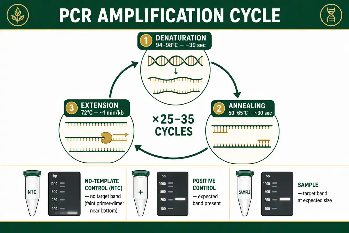
NCBI Bookshelf describes PCR as a laboratory nucleic acid amplification technique that uses DNA polymerase and temperature cycling to amplify DNA after denaturation and primer annealing. See Polymerase Chain Reaction. Another NCBI Bookshelf chapter explains that PCR makes copies of a selected DNA segment and became a major tool for studying genomes. See Studying DNA.
A useful PCR protocol should state the template source, target region, primer sequences or primer source, expected product size, polymerase type, control reactions, cycling design, contamination safeguards, and verification method. It should also explain what a valid result looks like and what will count as a failed reaction.
Good PCR controls include a no-template control, positive control template, and sometimes an internal amplification control. Without controls, a band on a gel may be real, contaminated, nonspecific, or the wrong product.
Common PCR problems include poor DNA quality, inhibitors, primer-dimer formation, nonspecific amplification, wrong annealing conditions, too much template, too little template, polymerase mismatch, contamination, and overinterpreting a faint band without confirmation.
Agarose Gel Electrophoresis and Fragment Analysis
Gel electrophoresis protocols separate DNA fragments by size. In genetics, gels are commonly used to check PCR products, estimate fragment length, confirm restriction digests, compare genotyping patterns, screen cloning products, or teach DNA analysis.
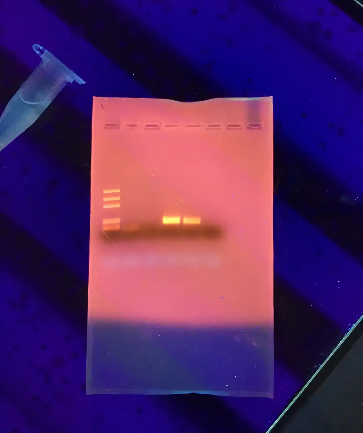
(Ultrabem, CC0, via Wikimedia Commons)
Health Canada describes gel electrophoresis as a basic technique used to separate DNA, RNA, or proteins. See Molecular techniques in biotechnology, science and research.
A useful gel electrophoresis protocol should state the gel concentration, buffer, DNA ladder, sample loading plan, stain, voltage or run condition, expected band size, imaging method, and interpretation rule. The ladder matters because it lets the reader compare unknown fragments to known sizes.
A gel can show whether a fragment is present and roughly how large it is, but it does not prove sequence identity by itself. For many genetics workflows, a gel is a screening or confirmation step, not the final interpretation.
Restriction Digestion and RFLP Analysis
Restriction fragment length polymorphism, or RFLP, uses restriction enzymes to cut DNA at specific recognition sequences. If a genetic variant creates or removes a restriction site, digestion can produce a different fragment pattern after gel electrophoresis.

RFLP is an older but still educationally useful genetics method. It helps students see how a sequence difference can become a visible banding pattern. It has also been used historically in mapping, genetic testing, strain comparison, and forensic genetics.
A useful restriction digestion or RFLP protocol should state the DNA template, enzyme, recognition site, expected fragment sizes, incubation condition, control digestion, undigested control, gel system, and interpretation rule. Partial digestion can create misleading bands, so the protocol should explain how complete digestion will be checked.
Genotyping and Marker Analysis
Genotyping protocols determine which allele, marker, or variant is present in a sample. Genotyping may use PCR, allele-specific PCR, restriction digestion, fragment analysis, SNP arrays, TaqMan assays, melt-curve analysis, sequencing, or other methods.
Genotyping is used in plant breeding, animal breeding, mouse colony management, human genetics, conservation genetics, ancestry studies, microbial strain analysis, pharmacogenetics, and research on genetic association.
A useful genotyping protocol should state the loci, reference sequence or marker name, allele definitions, primer or probe design, control genotypes, expected result patterns, quality thresholds, repeat rule, and how uncertain calls will be handled.
Genotyping mistakes are often interpretation mistakes. A faint band, mixed sample, allele dropout, contamination, sample swap, poor cluster separation, or wrong reference allele can lead to a confident but wrong genotype call.
DNA Sequencing Methods
DNA sequencing protocols determine the order of nucleotides in DNA. Sequencing may be targeted to one gene, focused on a panel of genes, applied to an exome, or expanded to whole-genome sequencing.
The National Human Genome Research Institute explains that DNA sequencing determines the order of bases in DNA and that technology improvements have made sequencing faster, cheaper, and more routine than it was during the Human Genome Project era. See the NHGRI DNA Sequencing Fact Sheet.
Sanger sequencing is still useful for targeted sequence confirmation, small amplicons, plasmids, and validating specific variants. Next-generation sequencing, or NGS, is used when many DNA fragments need to be sequenced in parallel, such as targeted panels, exomes, genomes, microbial genomes, amplicon sequencing, or population-scale studies.
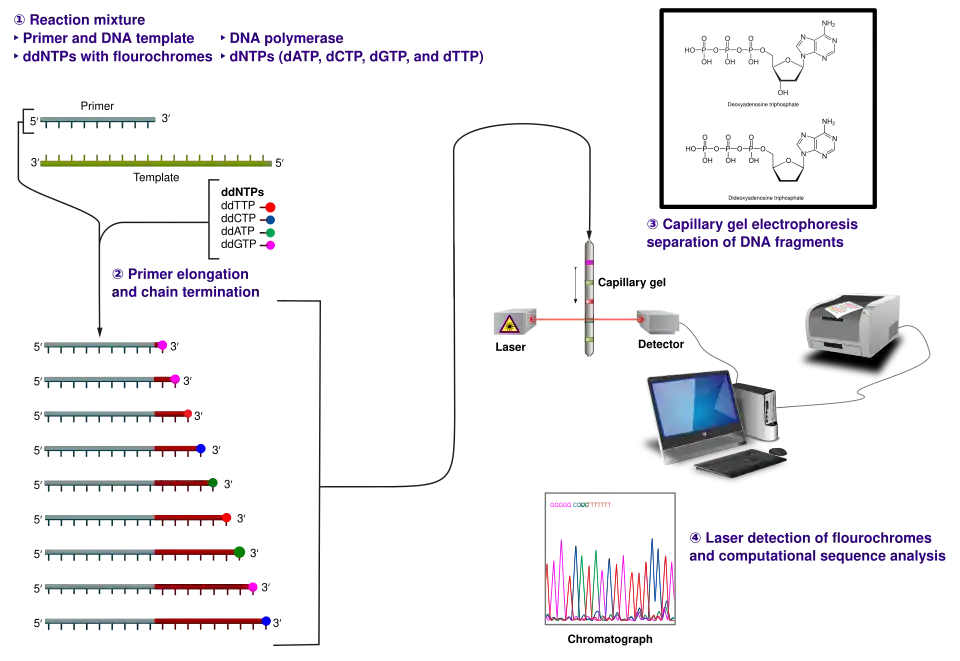
(Estevezj, CC BY-SA 3.0
A useful sequencing protocol should state the sample type, DNA input quality, library or amplicon strategy, target region, controls, indexing or barcoding method, sequencing platform, expected coverage, quality thresholds, reference genome, analysis workflow, and reporting limits.
Sequencing is powerful, but it does not automatically produce meaning. A variant may be real but harmless, real but uncertain, technical artifact, contamination, alignment error, or biologically important only in a specific context.
Next-Generation Sequencing Quality and Validation
Next-generation sequencing protocols need careful quality control because they combine wet-lab steps and computational analysis. A weak result can come from sample quality, library preparation, low coverage, index hopping, contamination, alignment errors, poor reference choice, or inappropriate filtering.
The CDC's NGS Quality Initiative describes a pathway to quality-focused testing that supports NGS assay validation, implementation, and maintenance because standardized quality frameworks are still challenging for NGS. See CDC Pathway to Quality-Focused Testing.
A useful NGS protocol should describe the assay type, specimen requirements, nucleic acid quality, library preparation, controls, sequencing metrics, coverage requirements, bioinformatics pipeline, variant filters, validation scope, and limitations. If results are used clinically, the laboratory must follow appropriate clinical-lab standards and reporting rules.
Research sequencing and clinical genetic testing are not the same thing. Research workflows can explore new questions. Clinical testing requires validation, quality systems, interpretation standards, and responsible reporting.
Variant Analysis and Interpretation
Variant analysis is the process of finding and interpreting DNA differences. These may include single-nucleotide variants, small insertions or deletions, copy-number changes, structural variants, repeat expansions, mitochondrial variants, or chromosomal abnormalities.
Variant interpretation depends on evidence. It may consider population frequency, predicted effect, segregation in families, functional data, disease association, inheritance pattern, phenotype match, and whether the variant has been reported before.
The CDC explains that large-scale genomic tests can include exome sequencing and whole genome sequencing, which look for genetic changes across a person's DNA and may be recommended when other testing has not found a genetic cause for a complex condition. See CDC Genetic Testing.
A useful variant-analysis protocol should state the reference genome, variant caller, quality filters, annotation tools, population databases, phenotype terms, inheritance model, classification system, confirmation plan, and reporting rules. It should also state how variants of uncertain significance will be handled.
For educational pages, this distinction is important: finding a variant is not the same as proving causation. Many DNA differences are benign, and some remain uncertain until more evidence is available.
RT-qPCR and Gene Expression Methods
RT-qPCR protocols measure RNA expression after RNA is converted into complementary DNA, or cDNA. In genetics and molecular biology, RT-qPCR is used to study gene expression, treatment response, developmental regulation, disease states, knockdown, overexpression, and pathway activity.
The MIQE guidelines were created to improve reliability, consistency, transparency, and reporting of quantitative real-time PCR experiments. See The MIQE guidelines.
A useful RT-qPCR protocol should state RNA extraction method, RNA quality, DNase treatment if used, reverse transcription conditions, primer design, amplification efficiency, reference genes, controls, replicate strategy, Ct or Cq handling, normalization method, and reporting approach.
Gene expression is not the same as genotype. Genotype describes DNA variation. Gene expression describes whether a gene is transcribed, and how strongly, under specific conditions. A genetics methods page should keep those ideas separate.
Pedigree Analysis and Inheritance Patterns
Pedigree analysis uses family relationships and trait patterns to infer possible inheritance modes. It is used in genetics education, medical genetics, animal breeding, plant breeding, and rare-disease investigation.
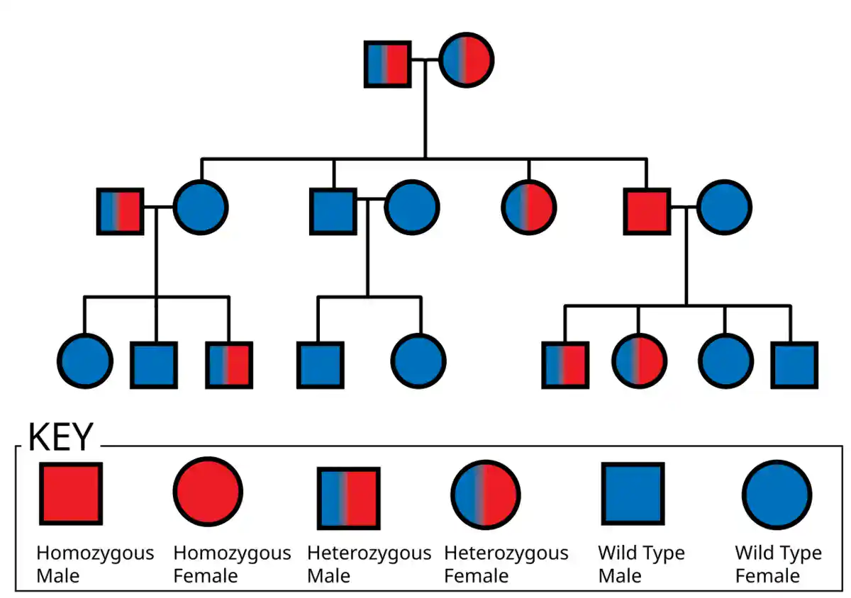
A useful pedigree-analysis protocol should state the trait definition, affected status criteria, relationship structure, sex of each individual, generation labels, consanguinity if relevant, known genotypes if available, penetrance assumptions, and uncertainty in the data.
Common inheritance patterns include autosomal dominant, autosomal recessive, X-linked dominant, X-linked recessive, Y-linked, mitochondrial, codominant, incomplete dominant, and multifactorial patterns. Real pedigrees can be complicated by incomplete penetrance, variable expressivity, de novo mutation, phenocopies, small family size, adoption, nonpaternity, mosaicism, or missing data.
For teaching and site strategy, this is where BioExplorer can connect naturally to the Punnett Square Calculator, genetics glossary, and pedigree analyzer.
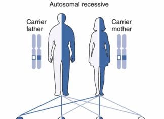
Autosomal Recessive Inheritance: Principles, Patterns, & Associated Disorders
Population Genetics Methods
Population genetics protocols study genetic variation across groups of individuals. These methods connect genetics with evolution, ecology, conservation biology, agriculture, epidemiology, ancestry, and species management.
Population genetics methods may estimate allele frequencies, genotype frequencies, heterozygosity, inbreeding coefficients, genetic differentiation, migration, effective population size, selection, drift, and Hardy-Weinberg expectations.
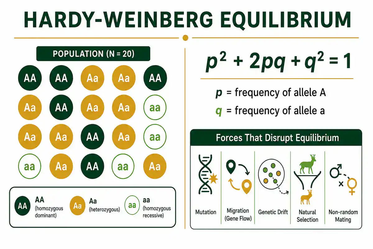
A useful population genetics protocol should state sampling design, population definition, marker type, genotyping method, sample size, missing data rule, allele-frequency calculation, Hardy-Weinberg test plan, correction for multiple testing if used, and interpretation limits.
Population genetics can be misread when sampling is weak. If individuals are not sampled representatively, if populations are mixed incorrectly, or if related individuals are overrepresented, allele-frequency estimates can become misleading.
CRISPR and Genome Editing Validation
Genome editing protocols are used to change DNA sequences or gene function in cells or organisms. CRISPR-based methods are widely used in genetics research to study gene function, create models, test regulatory sequences, engineer organisms, and validate candidate genes.
For a genetics methods hub, the most important point is validation. A claimed edit should be checked. Researchers often verify genome editing by PCR, gel electrophoresis, Sanger sequencing, targeted sequencing, restriction analysis, phenotype checks, protein assays, or off-target assessment depending on the experiment.
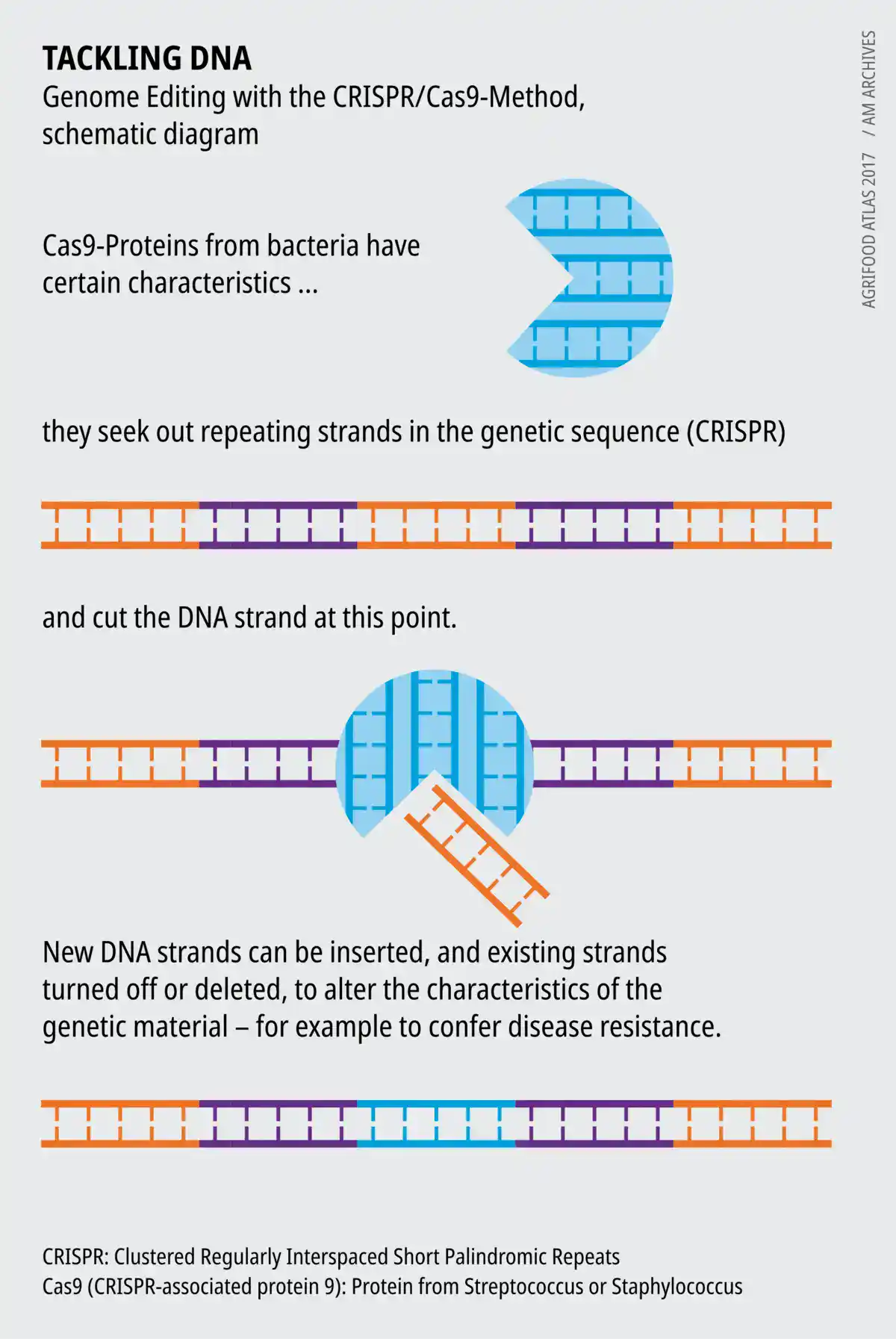
A useful genome-editing validation protocol should state the target region, guide design source, delivery approach, control group, genotyping method, expected edit, validation assay, mosaicism concerns, off-target strategy, and whether the result is clonal, pooled, somatic, germline, or transient.
This page should not turn genome editing into a casual recipe. The responsible focus is method selection, validation logic, controls, interpretation, safety, and ethics.
How to Choose the Right Genetics Protocol
Do not choose a genetics protocol only because it appears first in search results. Choose it because it matches your sample, question, target, required sensitivity, available equipment, safety rules, and interpretation plan.
Before using any genetics protocol, check these points:
- Sample type: blood, saliva, tissue, plant leaf, seed, microbial culture, environmental sample, forensic sample, ancient sample, or cultured cells.
- Question: extraction, amplification, mutation detection, inheritance, genotyping, expression, sequencing, population structure, or variant interpretation.
- Target: single gene, allele, SNP, STR, marker panel, transcript, exome, genome, mitochondrial DNA, or chromosome region.
- Method: PCR, gel electrophoresis, RFLP, Sanger sequencing, qPCR, RT-qPCR, SNP array, NGS, pedigree analysis, or population genetics analysis.
- Controls: positive control, negative control, no-template control, extraction blank, known genotype control, reference sample, or internal amplification control.
- Quality checks: DNA quantity, DNA purity, fragment integrity, PCR success, sequencing coverage, read quality, genotype call confidence, or replicate agreement.
- Interpretation: define how results will be called, confirmed, reported, and treated when uncertain.
- Ethics: human genetics, animal breeding, endangered species, clinical testing, ancestry, forensics, and gene editing may require special approval or caution.
Common Mistakes in Genetics Protocols
Genetics methods often fail for ordinary reasons. The technique may be valid, but the sample, control design, or interpretation may not match the question.
- Using poor-quality DNA: degraded or contaminated DNA can weaken PCR, sequencing, and genotyping results.
- Skipping extraction controls: contamination can enter before PCR or sequencing begins.
- Trusting PCR without controls: a band can be real, nonspecific, contaminated, or the wrong product.
- Overinterpreting gel bands: gel electrophoresis estimates size but does not prove exact sequence identity.
- Ignoring allele dropout: one allele may fail to amplify, especially in low-template or degraded samples.
- Mixing up genotype and phenotype: genotype describes genetic information, while phenotype is the observable trait or measurable outcome.
- Confusing gene expression with genotype: RNA levels do not necessarily tell you which DNA variant is present.
- Using weak reference genes in RT-qPCR: poor normalization can create false expression differences.
- Calling variants without context: a detected variant is not automatically disease-causing or biologically important.
- Ignoring population structure: allele-frequency comparisons can be misleading if populations are sampled or grouped poorly.
- Publishing incomplete methods: missing primer, reference genome, platform, analysis pipeline, or quality criteria makes results hard to interpret.
Genetics Calculators and Lab Tools
Genetics protocols often depend on calculations. A wrong dilution, primer design assumption, allele-frequency calculation, genotype ratio, Hardy-Weinberg estimate, qPCR fold change, or sequencing coverage estimate can affect the result before interpretation begins.
Useful genetics calculators include:
- Punnett square calculator
- monohybrid cross calculator
- dihybrid cross calculator
- blood type inheritance calculator
- Hardy-Weinberg equilibrium calculator
- allele frequency calculator
- genotype frequency calculator
- heterozygosity calculator
- inbreeding coefficient calculator
- DNA reverse complement calculator
- GC content calculator
- melting temperature calculator
- PCR master mix calculator
- DNA dilution calculator
- copy number calculator
- qPCR fold-change calculator
- sequencing coverage calculator
- variant allele frequency calculator
- pedigree probability calculator
- linkage map distance calculator
BioExplorer's Biology Tools and Calculators hub is building free browser-based tools by branch of biology. The Genetics and Inheritance Tools section is the best place to group genetics calculators.
Trusted Genetics Protocol Resources
Use BioExplorer as a guide, but always check your institution's approved SOPs, biosafety rules, ethics approvals, manufacturer instructions, and supervisor guidance before performing real laboratory work. These external resources are useful starting points for genetics protocols and method background:
- Nature Protocols: peer-reviewed protocol articles with design advice, troubleshooting, data analysis, limitations, and result interpretation.
- Current Protocols in Human Genetics: comprehensive methods resource for human genetics research and clinical laboratory approaches.
- NCBI Bookshelf: Polymerase Chain Reaction: background on PCR as a nucleic acid amplification technique.
- NCBI Bookshelf: Studying DNA: overview of DNA cloning, PCR, and recombinant DNA methods.
- NHGRI DNA Sequencing Fact Sheet: clear explanation of DNA sequencing and how sequencing technology has changed genetics.
- CDC Genetic Testing: overview of genetic and genomic testing, including exome and genome sequencing.
- CDC Pathway to Quality-Focused Testing: quality-focused framework for NGS assay validation, implementation, and maintenance.
- MIQE Guidelines: minimum information guidelines for reporting quantitative real-time PCR experiments.
- Health Canada Molecular Techniques: overview of molecular biology techniques including PCR and gel electrophoresis.
- Biology Methods and Protocols: Oxford Academic journal covering methods across biological research.
Safety, Ethics and Responsibility
Genetics protocols can involve human samples, animal samples, plant material, microorganisms, recombinant DNA, gene editing, clinical test results, ancestry data, forensic samples, endangered species, family information, personal identity, and medically sensitive findings. This page is educational. It does not replace formal training, institutional SOPs, biosafety approval, ethics approval, clinical laboratory rules, genetic counseling, or supervision by qualified personnel.
Human genetics deserves special care because genetic information can affect relatives, privacy, identity, medical decisions, and future risk interpretation. Clinical genetic testing should be performed and interpreted through qualified professionals and appropriate laboratory standards.
For gene editing, human-derived material, animal work, endangered species, forensic work, or clinical testing, follow approved institutional procedures and relevant legal, ethical, and biosafety requirements.
Related BioExplorer Resources
- Biology Methods and Protocols: main hub for biology lab methods, protocol categories, safety notes, and trusted resources.
- Genetics: guide to genes, DNA, heredity, chromosomes, mutations, inheritance, and genetic variation.
- Genetics Glossary: clear definitions of alleles, chromosomes, genotype, phenotype, DNA, mutations, inheritance, and related terms.
- Molecular Biology Methods and Protocols: methods for DNA, RNA, PCR, cloning, gel electrophoresis, sequencing prep, and molecular analysis.
- Biochemistry Methods and Protocols: methods for protein assays, enzyme kinetics, chromatography, western blotting, and biochemical analysis.
- Microbiology Methods and Protocols: methods for microbial culture, Gram staining, serial dilution, CFU counting, antimicrobial testing, and microbial molecular methods.
- Genetics and Inheritance Tools: browser-based tools for inheritance, DNA, and population genetics calculations.
- Punnett Square Calculator: predict genotype and phenotype probabilities for Mendelian crosses.
Frequently Asked Questions
Genetics protocols are written laboratory or analysis workflows for studying DNA, genes, chromosomes, variants, inheritance, genotypes, gene expression, pedigrees, and population-level genetic patterns.
Common genetics methods include DNA extraction, PCR, gel electrophoresis, genotyping, restriction digestion, Sanger sequencing, next-generation sequencing, RT-qPCR, pedigree analysis, variant analysis, and population genetics analysis.
Genetics focuses on heredity, genes, variation, inheritance, and genotype-phenotype relationships. Molecular biology focuses more broadly on DNA, RNA, proteins, gene expression, and molecular mechanisms inside cells.
PCR is used to amplify selected DNA regions so they can be checked, compared, sequenced, cloned, genotyped, or used in mutation detection and genetic analysis.
Genotyping is the process of determining which allele, marker, or DNA variant is present at one or more genetic locations in a sample.
No. Genotyping usually tests selected known variants or markers. DNA sequencing reads the order of DNA bases across a target region, panel, exome, genome, or other sequencing design.
A gel electrophoresis result shows DNA fragments separated mainly by size. It can confirm whether a PCR product or restriction fragment is present, but it does not prove the exact DNA sequence by itself.
Controls help distinguish real genetic results from contamination, failed amplification, nonspecific bands, poor DNA quality, sample swaps, allele dropout, sequencing artifacts, or analysis errors.
Not always. Online protocols vary in quality and may not match your sample, organism, equipment, biosafety level, ethics requirements, or interpretation goal. For real lab work, follow approved SOPs, safety guidance, ethics approval, and supervisor instruction.
Cite this page
BioExplorer. (2026, August 26). Genetics Methods and Protocols. https://www.bioexplorer.net/methods_and_protocols/genetics/