Do Prokaryotes Have Mitochondria?
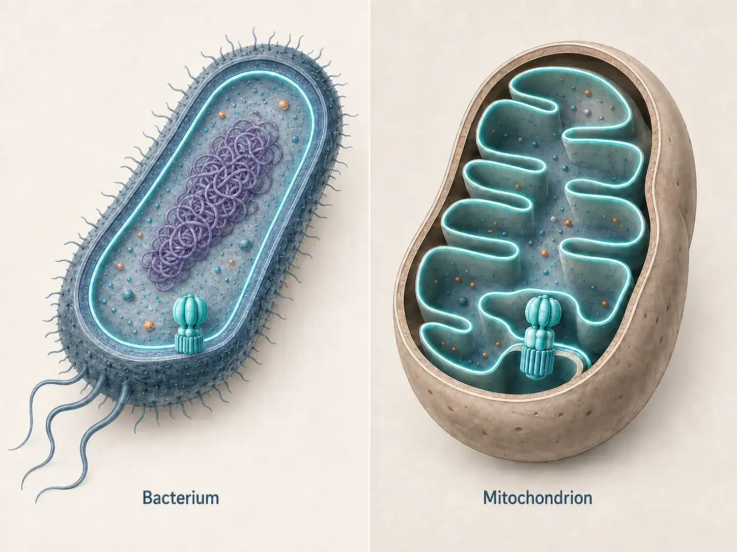
No. Prokaryotes do not have mitochondria. Neither bacteria nor archaea possess a mitochondrion, a nucleus, or any of the other canonical organelles that define eukaryotic cell architecture.
Prokaryotic cells nonetheless generate ATP. Respiring prokaryotes use chemiosmosis at a membrane the cell already possesses rather than at a dedicated organelle, whereas fermenters rely on substrate-level phosphorylation.
In most respiring bacteria that membrane is the plasma membrane. In most cyanobacteria and several other groups it is an internal membrane system.
The absence is not a deficiency. The mitochondrion is itself descended from a bacterium that became an endosymbiont within an archaeal host cell, which means the machinery of mitochondrial ATP synthesis originated in the prokaryotic world and was carried into eukaryotes rather than invented there.
Jump to:
- Summary of key facts
- What prokaryotes are
- Bacteria and Archaea are two separate domains
- The term describes an absence rather than a structure
- Prokaryotic DNA occupies a nucleoid
- How prokaryotes generate ATP without mitochondria
- Chemiosmosis operates at the plasma membrane
- ATP synthase types differ between the domains
- Cytosolic reactions and membrane reactions are distinct
- Three modes of energy conservation
- Nutritional classification of prokaryotes
- Sources of prokaryotic metabolic diversity
- Why prokaryotes lack mitochondria
- The membrane-area argument
- The energetic hypothesis is disputed
- Archaeal membranes solve the same problem differently
- The evolutionary origin of mitochondria
- Endosymbiotic theory
- The bacterial ancestor
- The archaeal host
- The order of events remains unresolved
- Two-domain and three-domain trees
- Evidence that mitochondria descend from bacteria
- An independent genome
- Non-Mendelian inheritance
- An independent translation system
- Bacterial machinery in the outer membrane
- The mitochondrial proteome is a mosaic
- The mesosome: a discredited bacterial organelle
- Magnetosomes
- The anammoxosome
- Thylakoids in cyanobacteria
- Carboxysomes and other protein-bounded microcompartments
- Eukaryotes that lack mitochondria
- Hydrogenosomes and mitosomes
- The amitochondriate oxymonads
- Prokaryotic cell and mitochondrion compared
- ❌ Common misconceptions
- FAQs
Summary of key facts
| Question | Answer |
|---|---|
| Do prokaryotes have mitochondria? | No. No bacterial or archaeal lineage has been found with one. |
| What performs the equivalent function? | The plasma membrane in most respiring species, or an internal membrane system in cyanobacteria and some others. |
| Where does glycolysis occur? | In the cytoplasm of prokaryotic cells. |
| Where does oxidative phosphorylation occur? | At a membrane, never free in the cytoplasm. |
| Do prokaryotes have any organelles? | Not the canonical eukaryotic set, but magnetosomes, anammoxosomes and thylakoids are genuine membrane-bounded compartments. |
| Where did mitochondria come from? | An alphaproteobacterial endosymbiont acquired by an archaeal host related to the Asgard archaea. |
| Are there eukaryotes without mitochondria? | Yes. Oxymonads such as Monocercomonoides exilis lost the organelle entirely. |
What prokaryotes are
Bacteria and Archaea are two separate domains
The term prokaryote covers two domains of cellular life: Bacteria and Archaea. These are deeply divergent lineages that differ in membrane chemistry, in cell envelope composition, and in the machinery of transcription and translation.
The distinction was established in 1977, when Carl Woese and George Fox compared 16S ribosomal RNA sequences and identified the archaea as a lineage separate from the bacteria. The proposal met sustained resistance and required roughly fifteen years to gain general acceptance.[1]
Defining prokaryotes as bacteria alone, as many introductory sources still do, omits an entire domain and obscures the archaeal ancestry of several eukaryotic systems.
The term describes an absence rather than a structure
Prokaryote is a negative definition. It specifies the features a cell lacks rather than the features it has, a point historians of biology have made in detail: the category was constructed on the basis of missing structures.[2]
The question of whether prokaryotes have mitochondria is therefore answered in part by the word itself. The substantive question is what performs the equivalent function.
Prokaryotic DNA occupies a nucleoid
Prokaryotic chromosomal DNA is not enclosed by a nuclear envelope. Neither is it free or disordered. It occupies a defined region called the nucleoid, where it is compacted and spatially organized by nucleoid-associated proteins. Further detail appears on the BioExplorer page covering whether bacteria have a nucleus.
How prokaryotes generate ATP without mitochondria
Chemiosmosis operates at the plasma membrane
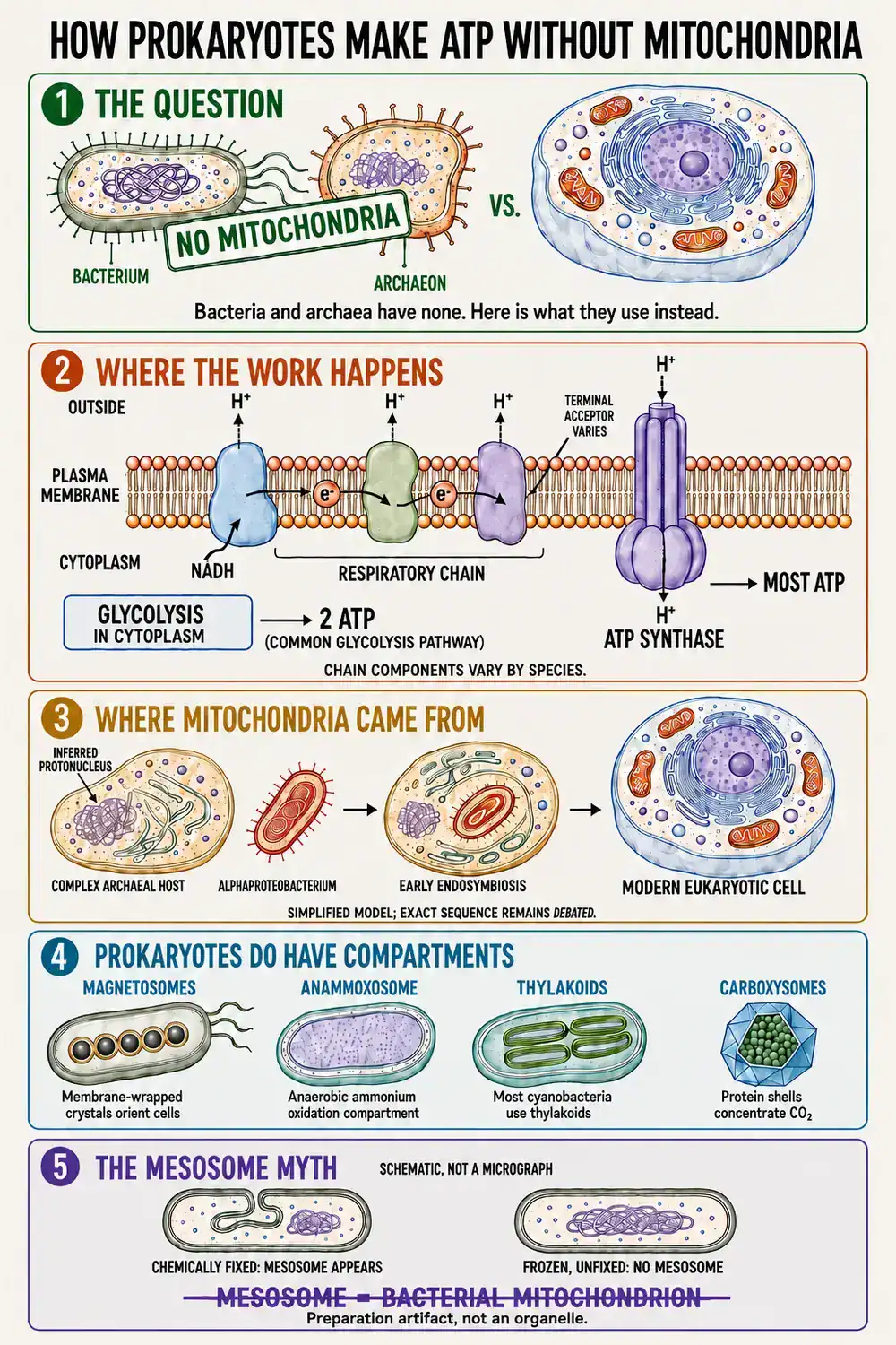
In a mitochondrion the functionally critical surface is the inner membrane, which carries the electron transport chain and the ATP synthase. Electrons pass along the chain, protons are pumped across the membrane, and the resulting electrochemical gradient drives ATP synthesis as protons return through the ATP synthase. The mechanism is chemiosmosis.
Respiring bacteria carry out the equivalent process at the plasma membrane. The standard reference description states that many bacteria oxidize sugars “by glycolysis, the citric acid cycle, and a respiratory chain in their plasma membrane that is similar to the one in the inner mitochondrial membrane“, and further that “the plasma membrane of the vast majority of bacteria contains an ATP synthase that is very similar to the one in mitochondria“[3]
ATP synthase types differ between the domains
The rotary ATP synthase family comprises three related classes namely, designated A-type, V-type and F-type. Bacteria typically use F-type enzymes and archaea typically use A-type or V-type enzymes, but the division is a tendency rather than a rule. The F-type family occurs in every domain of life, and exceptions in both directions are generally attributed to horizontal gene transfer.[4]
Ion coupling also varies. The proton motive force is the general case, but a number of archaea conserve energy using a sodium gradient instead, and archaeal bioenergetics accommodates both.[5]
Cytosolic reactions and membrane reactions are distinct
A frequent error in secondary sources is the statement that prokaryotes produce energy in the cytoplasm. The claim is imprecise rather than simply wrong, and the distinction is worth stating carefully.
Glycolysis, the oxidation of pyruvate, and most reactions of the citric acid cycle are soluble cytosolic processes in a bacterial cell. Glycolysis by the Embden-Meyerhof pathway yields a net two ATP per glucose through substrate-level phosphorylation, and alternative prokaryotic routes such as the Entner-Doudoroff pathway yield less.
What is not cytosolic is the terminal stage. The enzymes of electron transport and oxidative phosphorylation “reside on the bacterial inner (cytoplasmic) membrane”, and this is where the large ATP yield is generated.
The same reference records the range of terminal electron acceptors available under anaerobic conditions, including nitrate, sulfate, fumarate and carbon dioxide, and describes fermentation as the alternative in which an organic compound serves as the terminal acceptor.[6]
Three modes of energy conservation
- Aerobic respiration: Oxygen serves as the terminal electron acceptor. This mode generally supports the highest yields, although the figure depends on the electron donor, the acceptor and the architecture of the respiratory chain.
- Anaerobic respiration: A compound other than oxygen accepts the electrons. Nitrate, sulfate, fumarate and carbon dioxide are all used, according to the organism and its environment.
- Fermentation: No electron transport chain is involved. An organic molecule accepts the electrons, and the cell subsists on the modest yield of substrate-level phosphorylation.
The claim that mammalian cells are incapable of anaerobic respiration overstates the contrast. Under oxygen limitation, mammalian mitochondria can reverse the succinate dehydrogenase complex and deposit electrons onto fumarate, which then functions as an alternative terminal electron acceptor and sustains other mitochondrial activities.[7]
The accurate distinction is one of range. Mammals have a narrow known repertoire of respiratory electron acceptors; prokaryotes collectively use a wide one. The aerobic case is set out in full on the BioExplorer page covering the cellular respiration equation.
Nutritional classification of prokaryotes
Prokaryotic metabolic types are described by three independent variables. The compound terms used in the literature are these three answers combined.
| Variable | First option | Second option |
|---|---|---|
| Energy source | Phototroph: light | Chemotroph: chemical bonds |
| Electron source | Lithotroph: inorganic donors such as H2S or Fe2+ | Organotroph: organic donors |
| Carbon source | Autotroph: CO2 | Heterotroph: organic compounds |
Many cyanobacteria grow as photolithoautotrophs. Humans and Escherichia coli growing on sugar are both chemoorganoheterotrophs.
Sources of prokaryotic metabolic diversity
Prokaryotic metabolic versatility is sometimes attributed to the absence of organelles. That explanation does not hold. The diversity reflects the antiquity and breadth of prokaryotic lineages, extensive horizontal gene transfer, and the chemically extreme habitats many species occupy, where an unusual electron donor or acceptor may be the only one available.
Morphological and physiological variation across bacterial cell types reflects the same underlying diversity.
Why prokaryotes lack mitochondria
The membrane-area argument
The conventional explanation is geometric. Chemiosmosis requires membrane area, and the ratio of surface area to volume falls as cell size increases, because volume scales with the cube of the linear dimension while surface scales with the square.
Internalizing respiratory membrane within an organelle, and folding that organelle’s inner membrane into cristae, restores the available area. On this reasoning the mitochondrion addresses a constraint that only large cells encounter, and a bacterium one to a few micrometers across does not encounter it.
The energetic hypothesis is disputed
A stronger claim is often built on the geometric argument: that mitochondria conferred a decisive energetic advantage which made eukaryotic complexity possible. This claim is contested.
Analyses of how energy demand scales with cell volume have found continuous scaling across prokaryotes and eukaryotes rather than a discontinuity, and a subsequent quantitative treatment concluded that prokaryotes are not surface-area constrained across the volume range in which the earliest eukaryotes plausibly fell.
That analysis concluded that mitochondria may not have been required by the first eukaryotes, while eukaryotic diversification was ultimately dependent upon them.[8]
The defensible position is therefore layered. Membrane area is a genuine constraint on large, rapidly dividing cells. The proposition that mitochondria lifted an energy ceiling and thereby enabled cellular complexity remains a leading hypothesis rather than an established result.
Archaeal membranes solve the same problem differently
Archaea conserve energy chemiosmotically using membranes of a different chemistry. Archaeal membrane lipids are typically composed of isoprenoid chains joined by ether bonds to sn-glycerol-1-phosphate, whereas bacterial lipids typically consist of fatty acids joined by ester bonds to sn-glycerol-3-phosphate.
The two backbones are stereochemical mirror images with different linkage chemistry, a division regarded as one of the deepest in cellular biology, although exceptions to the clean separation are known.[9]
The evolutionary origin of mitochondria
Endosymbiotic theory
Endosymbiotic theory holds that mitochondria and plastids originated as free-living bacteria that were internalized by another cell and retained. The classical formulation states that the energy-converting organelles of eukaryotes “evolved from procaryotes that were engulfed by primitive eucaryotic cells and developed a symbiotic relationship with them”.[10] The characterization of the host as a eukaryotic cell is the element most affected by subsequent work.
Attribution requires care. Lynn Margulis, publishing under the name Lynn Sagan, presented the modern case in 1967 and defended it for decades against considerable resistance. She was not its originator. Constantin Mereschkowsky advanced a comparable proposal for chloroplasts in 1905, and Ivan Wallin argued for a bacterial origin of mitochondria during the 1920s. Margulis assembled the supporting evidence and sustained the hypothesis through a long period of rejection, which is a distinct contribution from originating it.
The bacterial ancestor
The mitochondrial endosymbiont derived from an alphaproteobacterial lineage. Comparative analysis of the mitochondrial proteome identified a core of proteins “descended from the ancestral alpha-proteobacteria symbiont”, while also establishing that the modern organelle is a mosaic whose proteins derive from several distinct sources.[11]
The precise relationship of the mitochondrial ancestor to extant alphaproteobacterial lineages is unresolved. Some analyses place it within the group and others recover it as a sister lineage to the sampled or core Alphaproteobacteria.
Phylogenetic approaches have produced inconsistent results depending on the method, the taxonomic sampling and the corrections applied for artifacts, and the identity of the closest living relatives is treated as an open question in the current literature.[12]
The archaeal host
The host cell was an archaeon. Phylogenomic analysis of an expanded sampling of Asgard archaeal genomes placed eukaryotes with high confidence as a well-nested clade within the Asgard archaea, as a sister lineage to the Hodarchaeales, a proposed order within the Heimdallarchaeia.[13]
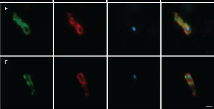 Hodarchaeales-related Asgard archaeal cells from Aarhus Bay sediment, imaged by CARD-FISH and super-resolution confocal microscopy. The first two columns show ribosomal RNA probe signal, the third shows DNA stained with DAPI, and the fourth is the overlay. Note the elongated cell shape and the single compact DNA focus. Image by Avcı et al., mBio, 2025, cropped from Figure 2. Licensed under CC BY 4.0.
The degree of cellular complexity the host had already achieved is the subject of active work. A relaxed molecular-clock analysis dated duplication events in gene families associated with eukaryotic cellular systems and inferred that substantial cytoskeletal, membrane-trafficking, endomembrane and nuclear or protonuclear complexity predated mitochondrial endosymbiosis. The timing of phagocytosis specifically is less certain: dates associated with the endolysosomal system overlap the earliest mitochondrial dates, and the authors state that they cannot discriminate which came first.
These duplication ages constitute maximum-age constraints on paralogue-specific functions rather than direct dates for the cellular structures themselves, and no known extant lineage represents the intermediate stages.[14]
Direct observation of Asgard archaea is consistent with a structurally complex host. Imaging of Asgard cells recovered from marine sediment identified cells up to approximately 5 micrometers in length with irregular morphology and confined central DNA, which the authors suggest could represent a transitional stage toward eukaryotic complexity.[15]
The order of events remains unresolved
Models span mitochondrion-early, mitochondrion-intermediate and mitochondrion-late scenarios, and the intermediate class is a substantive position rather than a midpoint between the other two.
At the two ends of this range, mitochondrion-late models hold that an already complexified archaeal host acquired the endosymbiont, while mitochondrion-early models hold that the endosymbiont preceded and financed the development of eukaryotic complexity.
The principal argument for the early position is energetic: a prokaryotic host lacking mitochondria and attempting to subsist by ingesting other prokaryotes would face a severe ATP deficit, on one estimate requiring the ingestion of approximately 34 times its body weight in prokaryotic prey per division, from which it follows that phagocytosis becomes physiologically viable only in a cell that already possesses mitochondria.[16]
The position supported by current evidence is limited but secure at its core. The host belonged to an archaeal lineage related to the Asgard archaea. Its precise degree of eukaryotic complexity, and the mechanism by which the endosymbiont was internalized, remain disputed. Describing the host as a proto-eukaryotic archaeal cell is defensible; describing it as a modern-style eukaryotic cell is not.
Two-domain and three-domain trees
Placing eukaryotes within the Asgard archaea implies a two-domain tree of life, which stands in tension with the three-domain framework in which Eukarya forms an independent branch. The element of Woese’s work that remains secure is the separation of Archaea from Bacteria. What has changed is the attachment point of Eukarya.
Improved evolutionary models and denser archaeal sampling now provide strong support for eukaryotes branching from within Archaea. Dissenting analysis published in 2018 raised objections concerning contamination in Asgard genome assemblies, the presence of conflicting phylogenetic signals within the same concatenated marker sets, and topologies that shift according to which fast-evolving taxa are included.[17]
Subsequent evidence has strengthened the two-domain position, and those objections are best understood as an earlier stage of the debate rather than a currently equivalent alternative.
Evidence that mitochondria descend from bacteria
An independent genome
Mitochondria retain their own genome. The human mitochondrial genome is a circular DNA molecule of 16,569 nucleotides, present in multiple copies per cell.[18]
Circularity should not be generalized. Investigation of plant mitochondrial DNA using fluorescence microscopy and pulsed-field gel electrophoresis found genome-sized circles to be exceedingly rare, with branched linear structures the most frequently observed physical form.[19] Mitochondrial genomes across eukaryotes may be circular, linear, branched or divided among multiple chromosomes.
Non-Mendelian inheritance
In humans and most other animals, mitochondria and mitochondrial DNA are inherited from the mother, which is the basis for using mitochondrial DNA to trace maternal lineages.[20] Other eukaryotic groups show biparental and paternal transmission. The animal pattern can be explored using the Mitochondrial Inheritance Calculator.
An independent translation system
Mitochondria maintain their own ribosomes. Human mitochondria retain mitoribosomes that synthesize the 13 proteins encoded by human mitochondrial DNA. These mitoribosomes have nevertheless diverged substantially from their bacterial origin, incorporating 36 mitochondria-specific proteins and losing several bacterial ribosomal elements.[21]
Descent from bacteria is well established; structural identity with bacterial ribosomes is not.
The figure of 13 protein-coding genes applies to humans and most other animals rather than to mitochondria generally. The set of genes retained in mitochondrial DNA, together with its organization and physical structure, varies markedly across eukaryotes.[22]
Bacterial machinery in the outer membrane
The mitochondrial inner membrane derives from the plasma membrane of the bacterial endosymbiont, which is why chemiosmosis occurs there. The outer membrane is not simply a residual host vesicle.
It contains beta-barrel proteins inserted by a dedicated sorting and assembly machinery whose core subunit, Sam50, belongs to the Omp85 superfamily and is homologous to the bacterial BamA subunit of the beta-barrel assembly machinery. The endosymbiotic origin of mitochondria is the accepted explanation for that conservation.[23]
The mitochondrial proteome is a mosaic
The small number of mitochondrially encoded proteins is sometimes read as a measure of how much of the endosymbiont genome relocated to the nucleus. That inference is unsound.
A proportion of ancestral bacterial genes was transferred, a substantial proportion was lost outright, and a large share of the modern mitochondrial proteome originated elsewhere: in the host lineage, in other bacteria, or as later eukaryotic innovations.
The mesosome: a discredited bacterial organelle
The generalization that prokaryotes possess no membrane-bounded organelles is a serviceable approximation at introductory level and inaccurate beyond it. Several well-characterized exceptions exist. None is a mitochondrion, and none is evolutionarily related to one.
Magnetosomes
Magnetotactic bacteria assemble chains of magnetite crystals that align the cell along geomagnetic field lines. Each crystal is enclosed within a membrane vesicle formed by invagination of the cytoplasmic membrane.
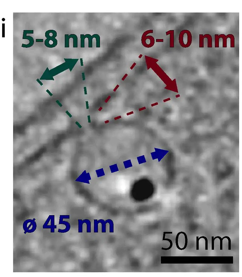
Magnetosomes are described in the literature as unique organelles and as among the most complex structures found in prokaryotic cells.[25] They constitute genuine lipids-bilayer-bounded organelles within a bacterium.
The anammoxosome
Anaerobic ammonium-oxidizing bacteria contain a large intracytoplasmic compartment, the anammoxosome, occupying most of the cell volume. It has been characterized as a prokaryotic organelle analogous to the eukaryotic mitochondrion, with the anammox reaction coupled across its curved membrane, possibly generating a proton motive force and supporting ATP synthesis.[26]
Its membrane contains ladderane lipids, which occur nowhere else in nature. The relationship to mitochondria is one of analogy rather than homology.
Thylakoids in cyanobacteria
Most cyanobacteria maintain an internal thylakoid membrane system, distinct from the plasma membrane and carrying a different protein complement. In these species the thylakoid membrane is the sole site of photosynthetic electron transport and the major site of respiration, while the plasma membrane carries transporters, sensors and components of the motility apparatus.[27]
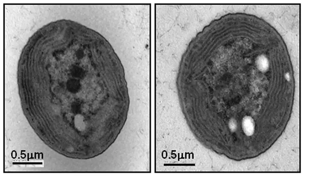
The arrangement is not universal. Members of the early-branching Gloeobacterales lack thylakoid membranes and conduct photosynthesis at the cytoplasmic membrane, a feature relevant to reconstructing the origins of oxygenic photosynthesis.[28]
A thylakoid is not a mitochondrion. The reason is not the absence of a dedicated genome, since many accepted organelles including lysosomes lack genomes. It is that the thylakoid differs from the mitochondrion in structure, in function and in evolutionary history.
Carboxysomes and other protein-bounded microcompartments
Carboxysomes enclose ribulose-bisphosphate carboxylase and carbonic anhydrase within a polyhedral shell, elevating local CO2 concentration and suppressing competing photorespiration. The shell is constructed entirely from protein. As one recent review states, “in contrast to the membrane-bound organelles of eukaryotes, prokaryotes utilize protein-based complexes to compartmentalize these reactions”.[29]
Prokaryotic compartmentalization is therefore real, but it does not always employ lipid membranes.
Eukaryotes that lack mitochondria
Hydrogenosomes and mitosomes
Numerous anaerobic eukaryotes carry reduced derivatives of the mitochondrion, known collectively as mitochondrion-related organelles. Hydrogenosomes generate ATP and release molecular hydrogen. Mitosomes do not generate ATP, and their retained functions vary between lineages, with iron-sulfur cluster assembly the most widespread.
As of a comprehensive 2012 review, no exception to the general rule was known: “All known eukaryotic groups possess an organelle of mitochondrial origin, mapping the origin of mitochondria to the eukaryotic common ancestor.”[30]
Variation between mitosomes is considerable. The mitosome of Entamoeba histolytica lacks the majority of recognized mitochondrial components and instead retains enzymes for sulfate activation, alongside lineage-specific proteins that make it markedly divergent from the mitosomes of other anaerobic protozoa.[31]
The amitochondriate oxymonads
The 2012 generalization no longer holds without exception. Monocercomonoides exilis, an oxymonad flagellate of the animal gut, was described as the first eukaryote arguably without any mitochondrion.
Subsequent genomic analysis confirmed complete mitochondrial loss in three oxymonad species, M. exilis, Blattamonas nauphoetae and Streblomastix strix, and concluded that the amitochondriate condition is likely common to the Oxymonadida as a whole, dating to a diversification approximately 100 million years ago.[32]
These organisms represent secondary loss rather than primitive absence. Their ancestors possessed mitochondria and dispensed with them after acquiring a replacement pathway for iron-sulfur cluster biosynthesis.
All current evidence on mitochondria and mitochondrion-related organelles remains consistent with a single ancestral acquisition.
Prokaryotic cell and mitochondrion compared
The following comparison describes typical cases. Most rows admit documented exceptions, noted beneath the table.
| Feature | Prokaryotic cell | Mitochondrion |
|---|---|---|
| Electron transport | Plasma membrane in most respiring species; internal membranes in cyanobacteria and some others; absent in fermenters | Inner membrane |
| ATP synthase | Typically F-type in bacteria, typically A-type or V-type in archaea, with exceptions in both directions; proton or sodium coupling | F-type, closely related to the bacterial enzyme |
| Soluble metabolism | Glycolysis, pyruvate oxidation and most of the citric acid cycle in the cytoplasm | Citric acid cycle in the matrix; glycolysis remains in the host cytosol |
| Genome | Usually a single circular chromosome in a nucleoid; linear and multiple chromosomes also occur | Own genome; 16,569 bp in humans, with structure and gene content varying widely across eukaryotes |
| Ribosomes | Own. Bacterial and archaeal ribosomes are distinct, and archaea share a set of ribosomal proteins with eukaryotes that bacteria lack | Own mitoribosomes, of bacterial descent but substantially modified |
| Membranes | A single plasma membrane in monoderms; an inner and an outer membrane in diderms such as gram-negative bacteria | Two, outer and inner |
| Propagation | Usually binary fission; budding and other modes also occur | Propagates through growth and fission; many mitochondria also undergo fusion; dynamics are host-controlled |
| Independent existence | Common, though obligate symbionts and intracellular parasites cannot survive independently | No. Most of its proteins are encoded in the host nucleus |
Gram-negative bacteria are diderms. Their envelope comprises a plasma membrane, a thin peptidoglycan layer and an outer membrane, with the periplasm occupying the intervening space, an architecture that distinguishes them from gram-positive cells and their thicker single-barrier walls.[33]
Binary fission is prevalent but not universal. Within the Planctomycetota, members of the class Planctomycetia reproduce by asymmetric budding while other classes divide by binary fission, and at least one described species initiates division by lateral budding along the side of the cell rather than at a pole.[34]
On ribosomes, a defined set of ribosomal proteins is present only in archaeal and eukaryotic ribosomes, contributing to the stabilization of ribosomal structure and to the interaction between subunits.[35] This is among the clearer structural indications of shared ancestry between the two groups.
❌ Common misconceptions
- That prokaryotes produce energy in the cytoplasm. Soluble metabolism is cytosolic. Electron transport and oxidative phosphorylation take place at a membrane.
- That prokaryote is a synonym for bacterium. The term covers Bacteria and Archaea, which differ in membrane chemistry, ATP synthase type and information-processing machinery.
- That a modern eukaryotic cell engulfed a bacterium. The host was an archaeon related to the Asgard archaea. Its complexity at the time, and the mechanism of internalization, remain disputed.
- That mesosomes are bacterial mitochondria. Mesosomes are an artifact of chemical fixation and are absent from living cells.
- That prokaryotes have no internal compartments. Magnetosomes, anammoxosomes, thylakoids and carboxysomes are all documented compartments.
- That all mitochondrial DNA is circular and encodes 13 proteins. Both claims describe human and typical animal mitochondria, not eukaryotes generally.
FAQs
No. No prokaryote, bacterial or archaeal, possesses a mitochondrion. Respiring species conduct electron transport at a membrane, most often the plasma membrane, using an ATP synthase closely related to the mitochondrial enzyme.
No. The relationship runs in the opposite direction: mitochondria are descended from bacteria. A bacterium has no mitochondrion because the mitochondrion is a derived, domesticated bacterium.
Pathways such as glycolysis can supply ATP by substrate-level phosphorylation. In respiring species, most ATP is generally generated by chemiosmosis at a membrane, where an electron transport chain establishes an ion gradient that ATP synthase converts into ATP. Terminal electron acceptors include oxygen in aerobic respiration and nitrate, sulfate, fumarate or carbon dioxide in anaerobic respiration. Fermenting species dispense with the electron transport chain entirely.
Chemiosmosis requires membrane area, and small cells have a high ratio of surface area to volume. The stronger claim, that mitochondria removed an energetic ceiling and thereby enabled cellular complexity, is a leading hypothesis rather than a settled conclusion, and quantitative analyses have found no clean energetic discontinuity between prokaryotes and eukaryotes.
Not the canonical eukaryotic set. The blanket statement that prokaryotes have no organelles is nonetheless too strong. Magnetotactic bacteria build membrane-bounded magnetosomes, anammox bacteria contain the anammoxosome, most cyanobacteria maintain thylakoid membranes, and many bacteria assemble protein-bounded microcompartments such as the carboxysome.
No. Cyanobacteria perform oxygenic photosynthesis using thylakoid membranes rather than a chloroplast, and the Gloeobacterales lack even those. The situation parallels mitochondrial evolution: primary plastids ultimately descend from cyanobacteria, whereas mitochondria descend from an alphaproteobacterial relative.
Yes, though very few. Oxymonads including Monocercomonoides exilis have lost the organelle completely. Many other anaerobic eukaryotes retain reduced derivatives in the form of hydrogenosomes or mitosomes. Every documented case represents loss from an ancestor that possessed mitochondria.
A mitochondrion cannot exist independently. Its genome encodes only 13 proteins in humans, and the remainder of its proteome is supplied by the host nucleus and drawn from several evolutionary sources. A free-living bacterium carries thousands of genes and reproduces autonomously.
Cite this page
BioExplorer. (2026, September 5). Do Prokaryotes Have Mitochondria?. https://www.bioexplorer.net/do-prokaryotes-have-mitochondria.html/
About the author

MSc Genetics & Cytology
Anastasiia Nesterenko holds an MSc in Genetics and Cytology and spent sixteen years as a research assistant at the Mechnikov Institute for Microbiology and Immunology. She has written for BioExplorer since 2016, from genetics and immunology to primates, insects, plants and flowers.