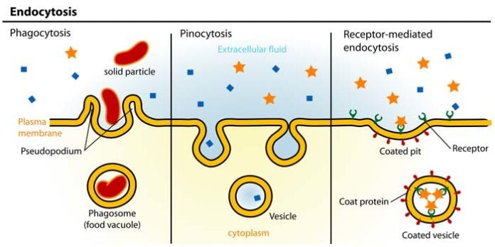
Cells are considered to be the basic unit of life and are mainly responsible for a wide variety of life processes.
Cells, through the course of evolution have adapted several ways to perform these tasks for survival. And did you know that like any other organisms, cells also need to breathe, eat, drink, and remove wastes?
In this article, you will learn about a certain process that somehow keeps the cell “hydrated” in order for it to continuously exist – pinocytosis.
Find out more below:
Table of Contents
What is Pinocytosis?
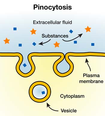 In cellular biology, pinocytosis is a type of endocytosis that involves the taking in of extracellular fluid into the cell.
- By utilizing this process, the cell can draw fluids and even solutes (as long as they are dissolved in fluids) into the cell with only a small amount of energy (ATP) needed. In particular, it is not the fluid itself that is needed by the cell but rather the molecules dissolved in it[1].
- When the cell engulfs fluid in, the fluid gets stored inside a vesicle (referred to as pinocytotic vesicle). Basically, this vesicle is a membrane-bound organelle that is made from the same lipid of the cell membrane.
![]()
The Discovery of Pinocytosis
- Amazed by how much this process appear like the cell drinking, Lewis called this phenomenon as “pinocytosis“, coming from the Greek word “pinos[2]” which means “I drink“.
- Three years later, this observation by Lewis was confirmed by scientists W.L.Doyle and S.O.Mast when they studied amoeba undergoing pinocytosis.
More Reading: W.H.Lewis (Proceedings of the American Association of Anatomists 78th Meeting – 1966). Search for “Warren Harmon Lewis“.
![]()
How Does It Work?
- To initiate the process, the cell membrane will have to allow the certain fluid it wants to engulf, resulting to an invagination in the structure.
- After which, the fluid continues to fill the invagination as it grows even larger to accommodate more fluid.
- The membrane then pinches off with the engulfed fluid trapped inside the vesicle.
- Basically, this entire process can be likened to the process offilling a balloon with air[3] . As you air, the balloon is filled before it is finally pinched off and tied, with the air trapped inside.
- Since the advent of electron microscopy, the process of pinocytosis was known to readily occur at different times in almost all types of cells.
The above video shows how a cell engulfs extracellular fluid into a vesicle and how this view looks like in an actual process (observed through Transmission Electron Microscopy). This was the original presentation by W.H.Lewis back in the last century.
![]()
Levels of Pinocytosis
There are apparently two distinguished levels of pinocytosis: micropinocytosis and macropinocytosis. The following are described below:
1. Micropinocytosis
| How to pronounce Micropinocytosis? | [responsivevoice buttontext=”Micropinocytosis”][/responsivevoice] |
As its name suggests, this level of pinocytosis occur in smaller scale. The vesicles formed in this pinocytosis have originated from the caveolae (depressions in the cell surface) and have a diameter of about 0.1µm.
- This process is highly insensitive to the inhibitors of microfilaments and may involve the protein[4] dynamin 2.
![]()
2. Macropinocytosis
| How to pronounce Macropinocytosis? | [responsivevoice buttontext=”Macropinocytosis”][/responsivevoice] |
In contrast to micropinocytosis, the vesicles formed during macropinocytosis are relatively larger with diameters ranging from 1 to 2µm. The vesicles originated from the invaginations of the surface ruffles or sometimes the plasma membrane.
- This is a type of pinocytosis exhibited by dendritic cells[5] in order to engulf fluid from the extracellular environment. This process helps them to take in antigens non-specifically.
![]()
Examples of Pinocytosis in Cells
As alluded to earlier, pinocytosis is a process that occurs almost all of the time. The following are some of the most common types of cells that perform pinocytosis:
1. Intestinal Cells With Microvilli
![]()
2. In Epithelial Cells
![]()
3. In Human Egg Cells
![]()
Difference Between Phagocytosis and Pinocytosis
The process of pinocytosis is often being related to phagocytosis with each of them having its own notable mechanisms and complexities. Tabulated below are the major differences between the two.
| PINOCYTOSIS | PHAGOCYTOSIS |
|---|---|
| Also known as “Cell-drinking“ | Also known as “Cell-eating“ |
| A type of endocytosis that involves the ingestion of fluids and solutes (dissolved in fluid) into the living cell. | A type of endocytosis that involves the engulfment of solid particles, foreign materials, and even other cells into the cell membrane. |
| The process can occur without the disruption of the cell’s cytoplasm. | May or may not involve the disruption of the cell’s cytoplasm (depending of the type of material to be ingested). |
| Smaller vesicles are formed. | Larger vesicles are formed. |
| No conversion is needed since the material is already dissolved and is ready for absorption. | Enzymes are needed to break down solid particles before they are allowed to enter the cell. |
Despite being induced by specific substances found in the extracellular environment, other materials like salt, minerals, and water can be found to be enclosed within the pinocytotic vesicles.
With this, scientists think that pinocytosis is predominantly nonspecific as compared with other types of endocytosis that are specific. An evidence for this is that all other molecules included in the fluid are taken into the cell.
On top of all of these, isn’t it amazing how such minute cell performs a very complicated task?
Cite This Page
References
- [1] – “avonapbio / endocytosis”. Accessed April 16, 2017. Link.
- [2] – “Types of Endocytosis: Pinocytosis, Receptor-Mediated Endocytosis and Phagocytosis”. Accessed April 16, 2017. Link.
- [3] – “Pinocytosis: Definition & Examples – Video & Lesson Transcript | Study.com”. Accessed April 16, 2017. Link.
- [4] – “Micropinocytosis.” Micropinocytosis – Oxford Reference. March 17, 2017. Accessed April 16, 2017. Link.
- [5] – “Search Results.” Oxford Reference. Accessed April 16, 2017. Link.
- [6] – “Pearson – The Biology Place”. Accessed April 16, 2017. Link.





very educative