Anatomy is the branch of biology that studies the physical structure of the human or animal body through direct observation, dissection, and microscopic examination of tissue. It ranks among the oldest branches of medicine, with study dating back to ancient Egypt and Greece.
This article traces that history from prehistoric cave art through modern tools like MRI, 3D bioprinting, and virtual dissection tables. The word comes from the Greek anatome, built from ana, meaning “up”, and tome, meaning “a cutting”, so it literally translates to “cutting up” or “dissection.”
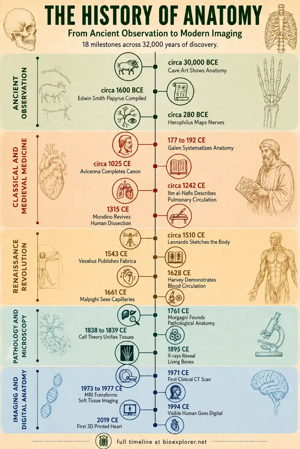
History of Anatomy timeline infographic showing 18 milestones from prehistoric cave art and the Edwin Smith Papyrus to Herophilus, Galen, Avicenna, Ibn al-Nafis, Leonardo da Vinci, Vesalius, Harvey, Malpighi, Morgagni, cell theory, X-rays, CT, MRI, the Visible Human Project, and the first 3D-printed heart.
See the Bigger Picture
Anatomy is just one thread in a much longer story. Follow the complete History of Biology to see how the study of body structure connects to genetics, evolution, and every discipline that came after it.
Prehistory (circa 30,000 to 3,000 BCE)
Circa 30,000 BCE:
The earliest known depictions of animal anatomy appear in cave art such as Chauvet Cave in France. The paintings show accurate musculature, proportion, and movement in animal figures, suggesting careful observation of living and hunted animals. There is no evidence yet of formal anatomical study, only close visual familiarity with animal bodies gained through hunting and butchering. (Source)
Ancient Egypt (circa 1600 to 1500 BCE)
Circa 1600 BCE:
The Edwin Smith Papyrus is compiled, though it is believed to be a copy of a much older Old Kingdom text. It describes 48 real surgical cases involving injuries, fractures, and wounds, with detailed observations of anatomy and a notably rational, non-magical approach to diagnosis and treatment. (Source)
Circa 1550 BCE:
The Ebers Papyrus is written, becoming the most extensive surviving record of Egyptian medical knowledge. Alongside magical remedies and hundreds of prescriptions, it shows real understanding of the heart and blood vessels, including the idea that the heart was central to a network of vessels carrying fluids through the body. (Source)
5th Century BCE (500 to 401 BCE)
Circa 500 BCE:
Alcmaeon of Croton performs some of the earliest recorded dissections of animals and possibly humans. He proposes that the brain, not the heart, is the seat of intelligence and sensation, a claim that put him at odds with the dominant thinking of his time. (Source)
Circa 450 BCE:
Empedocles introduces the concept of “pneuma“, describing it as a vital substance flowing through the blood vessels. His ideas blend early physiology with philosophy and influence Greek medical thought for generations afterward. (Source)
Circa 400 BCE:
Hippocrates, later called the Father of Medicine, argues that disease results from natural causes in the body rather than the will of the gods. He and his followers build a body of clinical observation, the Hippocratic Corpus, that ties symptoms to the body’s own processes rather than supernatural punishment. (Source)
4th Century BCE (400 to 301 BCE)
384 to 322:
Aristotle, working mostly through animal dissection, becomes known as the Father of Comparative Anatomy and Physiology. Some of his conclusions, such as the belief that the heart was the seat of intelligence and that arteries carried only air, were later proven wrong, but his comparative method across species shaped biological inquiry for centuries. (Source)
3rd Century BCE (300 to 201 BCE)
Circa 300 BCE:
The medical school at Alexandria is established under the early Ptolemaic rulers, creating a rare environment where human dissection, and for a brief period even vivisection of condemned criminals, was permitted. This openness would not be matched in the Western world again for roughly 1,500 years. (Source)
Circa 280 BCE:
CC BY 4.0, via Wikimedia CommonsHerophilus, working in Alexandria, documents at least 7 pairs of cranial nerves, distinguishes sensory from motor nerves, and identifies the heart valves. He also names parts of the eye still in use today, including the cornea and retina. (Source)
Circa 260 BCE:
Erasistratus builds on Herophilus’s work, describing the heart as a mechanical pump with distinct chambers and mapping the differences between veins and arteries. His anatomical drawings and descriptions mark one of the last major achievements of the short lived Alexandrian dissection era. (Source)
2nd Century CE (101 to 200 CE)
157 CE:
Galen becomes physician to the gladiators of Pergamum, gaining hands on knowledge of the human body through treating severe traumatic wounds. This role gives him an unusual, practical window into muscle, bone, and organ structure that shapes the rest of his career. (Source)
177 to 192:
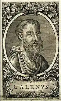
Galen writes On Anatomical Procedures, describing seven pairs of cranial nerves, arguing correctly that arteries carry blood rather than air, and demonstrating that urine forms in the kidneys rather than the bladder. His conclusions, some accurate and some mistaken since he could not dissect humans and relied heavily on animal anatomy, become the unquestioned standard for the next 1,300 years. (Source)
9th to 11th Century CE (801 to 1100 CE)
865 to 925:
Al Razi, known in the West as Rhazes, writes the Kitab al Hawi fi al Tibb, a 23 volume medical encyclopedia. It becomes core curriculum in European medical schools for centuries and preserves and extends Greek and Roman anatomical knowledge through the period when it had largely stalled in Europe. (Source)
936 to 1013:
Al Zahrawi, known as Albucasis, compiles the Kitab al Tasrif, a 30 volume medical text whose final section on surgery describes and illustrates dozens of surgical instruments, including scalpels, forceps, and cauterization tools he designed himself. He is often called the Father of Operative Surgery for the precision and detail of his anatomical and surgical descriptions. (Source)
Circa 1025 CE:
Avicenna completes the Canon of Medicine, a five volume work that systematizes anatomy, physiology, and treatment. Translated into Latin, it becomes a standard medical text in European universities well into the 17th century. (Source)
13th Century CE (1201 to 1300 CE)
Circa 1242 CE:
Ibn al Nafis, writing a commentary on Avicenna’s Canon, describes the pulmonary circulation, correctly arguing that blood travels from the right side of the heart through the lungs before reaching the left side, and that the wall between the heart’s chambers has no pores as Galen had claimed. His manuscript is lost to the West for centuries and only rediscovered in 1924, by which point Europe already credited the same discovery to William Harvey. (Source)
14th Century CE (1301 to 1400 CE)
1315 CE:
Mondino de Luzzi performs the first recorded public human dissection in Europe since antiquity, in Bologna, in front of medical students and other observers. The dissection, using the body of an executed criminal, was sanctioned by church authorities and marked the reintroduction of hands on human dissection to European medical education. (Source)
1316 CE:
Mondino completes the Anathomia, the first dedicated dissection manual and anatomical textbook of the modern era. It remains the standard European anatomy text for roughly 250 years. (Source)
16th Century CE (1501 to 1600 CE)
Circa 1510 CE:
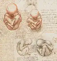
Leonardo da Vinci, having dissected around 30 human corpses over his lifetime, produces detailed anatomical sketches of the skeleton, muscles, brain, and organs, including an accurate drawing of a fetus attached by the umbilical cord. His drawings were not published in his lifetime and had little direct influence on the anatomy of his era, but they are now recognized as remarkably accurate for their time. (Source)
1543 CE:
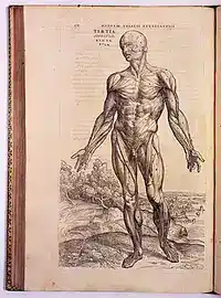
CC BY 4.0, via Wikimedia CommonsAndreas Vesalius publishes De Humani Corporis Fabrica in Basel, based on his own human dissections rather than inherited Galenic texts. The work corrects numerous errors in Galen’s centuries old descriptions and earns Vesalius the title Father of Modern Anatomy, though it also draws sharp criticism from anatomists loyal to Galen’s authority. (Source)
17th Century CE (1601 to 1700 CE)
1628 CE:
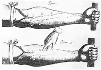
William Harvey publishes De Motu Cordis, demonstrating that blood circulates continuously through the body in one direction, driven by the heart acting as a pump. He could not see the capillaries connecting arteries to veins and described them only as an inferred “porosity“, a gap that would be closed decades later. (Source)
1661 CE:
Marcello Malpighi publishes De Pulmonibus, describing the capillaries in frog lungs under a microscope and confirming what Harvey could only theorize. This is the first direct observation of capillaries in any circulatory system. (Source)
1665 CE:
Robert Hooke publishes Micrographia, which includes the first recorded use of the word “cell” to describe the compartments he observed in cork tissue under a microscope. The book helps popularize microscopy as a serious scientific tool across Europe. (Source)
18th Century CE (1701 to 1800 CE)
1752 CE:
England passes the Murder Act, which allows the bodies of executed murderers to be legally dissected as an added punishment beyond hanging. Public dissections at scaffolds become part of the sentence, tying anatomical study to criminal justice in a way that shapes the supply of cadavers for the next 80 years. (Source)
1761 CE:
Giovanni Battista Morgagni publishes De Sedibus et Causis Morborum per Anatomen Indagatis, compiling more than 700 case histories paired with post mortem findings. The work founds pathological anatomy by systematically linking symptoms observed in life to physical changes found after death. (Source)
19th Century CE (1801 to 1900 CE)
1832 CE:
Britain passes the Anatomy Act, allowing medical schools to use unclaimed bodies for dissection and ending the practice of relying solely on executed criminals. The law responds directly to the body snatching scandals of the preceding decades, including the Burke and Hare murders in Edinburgh. (Source)
1838 to 1839:
Matthias Schleiden and Theodor Schwann jointly establish cell theory, concluding that all living tissue, plant and animal alike, is built from cells. This becomes a foundational principle for microscopic anatomy and histology going forward. (Source)
1858 CE:
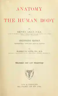
Henry Gray, with illustrations by Henry Vandyke Carter, publishes Anatomy Descriptive and Surgical. The two worked together for about 18 months, dissecting unclaimed bodies made available under the Anatomy Act, and the book, now known simply as Gray’s Anatomy, remains a core reference for medical students today. (Source)
1895 CE:
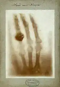
Wilhelm Rontgen discovers X-rays while experimenting with cathode ray tubes in his lab in Wurzburg. For the first time, the internal structure of a living body, bones in particular, can be seen without any incision, and the discovery earns him the first Nobel Prize in Physics in 1901. (Source)
20th Century CE (1901 to 2000 CE)
1901 CE:
Karl Landsteiner identifies the A, B, and O blood groups, explaining why some blood transfusions succeeded and others caused fatal reactions. The discovery makes safe blood transfusion possible and earns him the 1930 Nobel Prize in Physiology or Medicine. (Source)
1931 CE:
Ernst Ruska and Max Knoll build the first electron microscope, and within a few years Ruska’s team begins adapting the technology to examine biological specimens. This eventually opens up cellular and subcellular anatomy to direct observation at a resolution far beyond what light microscopes could achieve. (Source)
1971 CE:
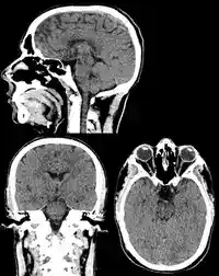
Godfrey Hounsfield performs the first clinical CT scan on a patient with a suspected brain tumor at Atkinson Morley Hospital in London. It is the first time a cross sectional image of a living brain is produced without surgery, and the technology is unveiled publicly the following year. (Source)
1973 to 1977:
Mikael Häggström, M.D. CC0, via Wikimedia CommonsPaul Lauterbur, Raymond Damadian, and Peter Mansfield each contribute key techniques that lead to magnetic resonance imaging, allowing detailed soft tissue images of the body without radiation. MRI becomes one of the most important tools in both clinical anatomy and research. (Source)
1994 CE:
The United States National Library of Medicine releases the Visible Human Project dataset, consisting of complete MRI, CT, and cross sectional photographic images of a human male cadaver. It becomes a widely used digital reference for anatomy education and remains influential in the development of 3D anatomical modeling. (Source)
21st Century CE (2001 to present)
2010 CE:
The National Institutes of Health (NIH) launches the Human Connectome Project, a large scale effort to map the structural and functional wiring of the healthy human brain using advanced imaging techniques. The project produces one of the most detailed maps of brain connectivity ever assembled and continues to inform research into neurological and psychiatric disorders. (Source)
2011 CE:
Anatomage releases the first life size virtual dissection table, combining CT and MRI scan data of real human bodies into an interactive touchscreen platform. It is adopted by medical, dental, and veterinary schools worldwide as a supplement to, and in some programs a partial replacement for, cadaver dissection. (Source)
2019 CE:
Researchers at Tel Aviv University 3D print the first heart containing human tissue, blood vessels, ventricles, and chambers, built from a patient’s own cells. The heart, roughly the size of a rabbit’s, does not yet function as a pump, but it is the first time a complete heart structure with vasculature has been engineered this way, marking an early step toward bioprinted transplant organs. (Source)
There were several advancements, breakthroughs, and innovations in several fields of biology in 2017. In this article, let’s glance over the top 15 anatomy and physiology related breakthroughs in 2017.
The advances in clinical medicine show us that our bodies still hold many secrets. Here are the top 26 anatomy and physiology news in 2018 (discoveries and breakthroughs).
From the scientific point of view, the year 2020 was devoted to public health, vaccine, and virus research. Here are top 10 anatomy/physiology news of 2020.
These 2021 noteworthy advancements and discoveries in Anatomy also speak to scientists’ increased interest in the brain’s structure, function, and disorders. Explore the top 15 anatomy and physiology news of 2021.
Anatomy/Physiology News of 2022: Discover breakthroughs in organ transplant technology, multi-organ chips, Alzheimer’s research, X-ray immunotherapy, astronauts’ brain studies, and more.
Anastasiia Nesterenko holds an MSc in Genetics and Cytology and spent sixteen years as a research assistant at the Mechnikov Institute for Microbiology and Immunology. She has written for BioExplorer since 2016, from genetics and immunology to primates, insects, plants and flowers.
Cite this page
BioExplorer. (2026, September 30). History of Anatomy. https://www.bioexplorer.net/history_of_biology/anatomy/
Highly impressive detailed development phase of anatomy. Thanks to all of the contributors since antiquity, without which I wouldn’t have been so vast in my discipline.






Highly impressive detailed development phase of anatomy. Thanks to all of the contributors since antiquity, without which I wouldn’t have been so vast in my discipline.
Perfectly explained ?