
Microbiology Protocols

Microbiology protocols are practical laboratory methods used to study bacteria, archaea, fungi, protists, viruses, microbial communities, and the conditions that affect microbial growth, survival, identification, and behavior. These methods help researchers culture microbes, prepare sterile media, isolate colonies, stain cells, count viable cells, test antimicrobial response, study biofilms, extract microbial DNA, and connect microbial results back to a biological question.
This page is a guide to major microbiology methods and protocols used in research labs, teaching labs, clinical microbiology, environmental microbiology, food microbiology, biotechnology, molecular biology, immunology, and public health. It explains what each method is for, what a good protocol should include, where mistakes usually happen, and which trusted resources can help you go deeper.
A useful microbiology protocol is more than a list of steps. It should explain the organism or sample type, biosafety requirements, culture conditions, media, controls, contamination checks, incubation variables, data readout, waste handling, troubleshooting, and limitations. The CDC's Biosafety in Microbiological and Biomedical Laboratories emphasizes protocol-driven risk assessment because laboratory risk depends on the agent, procedure, controls, equipment, facility, and worker competency.
What Are Microbiology Protocols?
Microbiology protocols are written workflows for handling, observing, growing, identifying, counting, or testing microorganisms under defined conditions. They may be used to culture bacteria, isolate single colonies, perform a Gram stain, count colony-forming units, test disinfectants, measure microbial growth, detect contamination, study biofilms, or prepare microbial DNA for molecular analysis.
The method is the scientific technique. The protocol is the practical workflow for using that technique. For example, serial dilution is a method for reducing microbial concentration in a stepwise way. A serial dilution protocol explains the dilution series, mixing method, plating method, controls, incubation conditions, plate-counting rules, and how colony counts will be converted into CFU/mL or CFU/g.
Microbiology overlaps closely with microbiology, molecular biology, biochemistry, immunology, biotechnology, ecology, medicine, epidemiology, food science, environmental science, and public health.
Microbiology Protocols Guide
Use this page as a map for common microbiology lab methods and microbial analysis workflows.
- biosafety and biological risk assessment
- aseptic technique and contamination control
- sterile media preparation
- bacterial culture and colony isolation
- streak plate, spread plate, and pour plate methods
- serial dilution and dilution theory
- CFU counting and viable plate counts
- Gram staining and basic microbial microscopy
- microbial growth curves and OD600 measurements
- antimicrobial susceptibility testing
- biofilm assays and crystal violet staining
- environmental and food microbiology sampling
- microbial DNA extraction and molecular identification
- controls, troubleshooting, and quality checks
Core Microbiology Methods at a Glance
The table below gives a quick map of important microbiology methods and what each one helps measure or control.
| Method | Main Purpose | Common Readout |
|---|---|---|
| Aseptic technique | Reduce contamination during microbial handling | Sterile cultures, clean plates, uncontaminated controls |
| Media preparation | Provide nutrients and selective conditions for growth | Liquid broth, agar plates, selective or differential media |
| Streak plate | Isolate single colonies from a mixed or dense sample | Separate colonies, colony morphology, pure culture selection |
| Spread plate | Distribute diluted cells across an agar surface | Countable colonies, CFU estimate, growth distribution |
| Serial dilution | Reduce microbial concentration step by step | Dilution series, countable plates, CFU/mL or CFU/g calculation |
| Gram stain | Differentiate many bacteria by cell wall staining behavior | Gram-positive or Gram-negative staining pattern, cell shape, arrangement |
| CFU counting | Estimate viable cells able to form colonies | Colony count, dilution factor, CFU/mL, CFU/g, log reduction |
| Growth curve | Track microbial growth over time | Lag phase, exponential phase, stationary phase, OD600, generation time |
| Antimicrobial susceptibility testing | Assess microbial response to antimicrobial agents | Zone of inhibition, MIC, susceptible/intermediate/resistant interpretation |
| Biofilm assay | Measure surface-attached microbial biomass or behavior | Crystal violet staining, absorbance, biofilm biomass, treatment effect |
Biosafety and Biological Risk Assessment
Microbiology protocols must begin with safety. Microorganisms are not interchangeable. A harmless classroom organism, a food isolate, an environmental sample, a clinical specimen, and a genetically modified strain can require very different containment, training, waste handling, and approval.
The CDC describes biological risk assessment as a process that identifies hazards, evaluates risks, implements controls, and reviews whether those controls are effective. The assessment should consider the biological agent, specific procedures, existing controls, facility, testing environment, and worker competency. See CDC Biological Risk Assessment.
A strong microbiology protocol should state the organism or sample category, required biosafety level, PPE, engineering controls, disinfection method, waste disposal method, spill response, aerosol-generating steps, sharps policy, and whether institutional approval is required. For unknown environmental or clinical samples, risk should not be guessed from appearance.
This page is educational. It is not a substitute for institutional SOPs, biosafety committee guidance, supervisor training, or organism-specific safety rules.
Aseptic Technique and Contamination Control
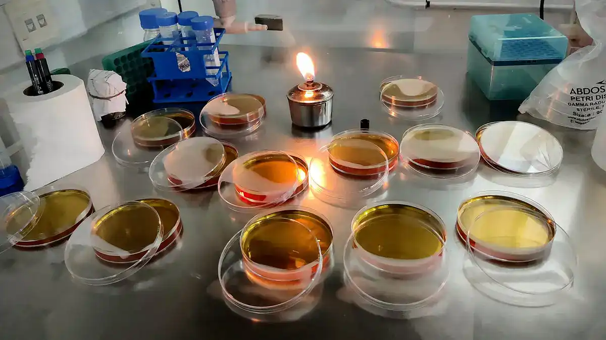
Aseptic technique is the foundation of microbiology lab work. It reduces the chance that unwanted microbes enter a culture, sample, reagent, plate, pipette tip, tube, or work surface. It also helps protect the worker, the experiment, and the environment.
A useful aseptic technique protocol should describe the work area, PPE, hand hygiene, sterile tools, tube and plate handling, flame or no-flame policy, sterile transfer technique, labeling, negative controls, waste handling, and cleanup. It should also explain how to recognize contamination and when to discard a culture or plate.
Plating methods are a common place where aseptic technique matters. A peer-reviewed JoVE article on plating methods describes streak plate, spread plate, and pour plate methods as aseptic laboratory techniques for isolating, spreading, and counting microorganisms on solid media. See Aseptic Laboratory Techniques: Plating Methods.
Common aseptic technique problems include leaving plates open too long, touching sterile tips to nonsterile surfaces, mixing up labels, working over crowded benches, using wet agar plates, failing to include negative controls, and assuming that a single clean-looking plate proves the whole workflow was clean.
Culture Media and Bacterial Culture Protocols
Microbial culture protocols describe how microorganisms are grown under defined conditions. Culture methods may use liquid broth, solid agar, selective media, differential media, enrichment media, anaerobic conditions, defined media, complex media, or specialized conditions depending on the organism and goal.
Culture media are not just food for microbes. Medium composition can select for some organisms, suppress others, reveal biochemical traits, or change growth rate. A culture protocol should state the medium, source, preparation method, storage conditions, sterility check, organism, inoculum source, incubation atmosphere, incubation time, temperature range if applicable, and expected colony or turbidity result.
A review on bacterial culture explains that solid media are prepared by adding a gelling agent such as agar to culture broth, allowing isolated colonies of different bacterial species to be obtained and described by morphology. See Bacterial culture through selective and non-selective conditions.
A good culture protocol also explains limitations. Many microbes are difficult to culture, grow slowly, require special nutrients, depend on other organisms, or change behavior outside their natural environment. Culture tells you what grew under the chosen conditions, not necessarily everything that was present in the original sample.
Streak Plate, Spread Plate and Pour Plate Methods
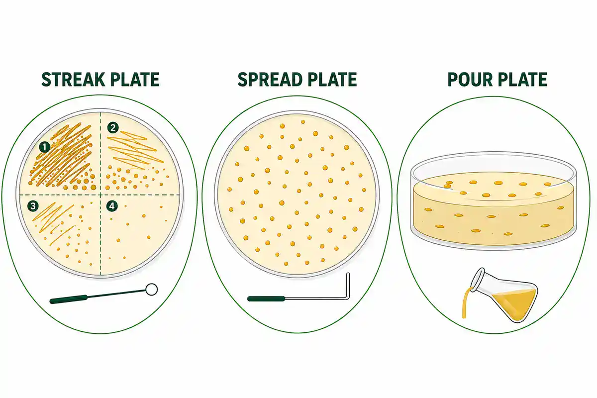
Plating protocols are used to isolate colonies, check purity, estimate viable cell numbers, compare treatments, or recover microbes from a sample. The most common methods are streak plate, spread plate, and pour plate.
- Streak plate: used mainly to dilute cells across the plate surface and isolate single colonies.
- Spread plate: used to spread a measured volume of diluted sample across an agar surface for colony counting or recovery.
- Pour plate: mixes sample with molten agar so colonies can form within and on the surface of the medium.
The spread plate method, often used with serial dilution, is valuable for quantitative work. A microbiology education article notes that spread plating with serial dilutions requires repeatable, reliable technique to produce meaningful results. See Perfecting Your Spread Plate Technique.
A useful plating protocol should specify plate type, dilution, plated volume, spreading method, drying conditions, controls, incubation conditions, colony-counting rule, and how contamination or confluent growth will be handled. If the goal is pure culture isolation, colony selection and restreaking rules should also be included.
Serial Dilution and CFU Counting Protocols
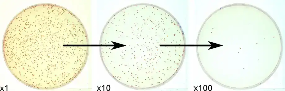
Serial dilution protocols reduce microbial concentration in a stepwise way so that at least one plated dilution gives a countable number of colonies. This is central to viable plate counts, microbial survival studies, disinfectant testing, food microbiology, environmental microbiology, fermentation, and antimicrobial research.
CFU counting estimates the number of viable cells or clumps capable of forming colonies under the chosen conditions. CFU does not always equal the exact number of individual cells because one colony can arise from a single cell or a cluster of cells.
The American Society for Microbiology's serial dilution protocol states that the standard plate count is a reliable method for enumerating bacteria and fungi and uses a set of serial dilutions followed by plating. See ASM Serial Dilution Protocols. A microbiology commentary also notes that many textbooks and lab manuals recommend using plates with roughly 30 to 300 colonies because plates with too few or too many colonies are harder to interpret reliably. See Plate Counting You Can Count On.
A useful CFU protocol should state the dilution scheme, mixing method, plated volume, number of replicate plates, incubation conditions, countable range, colony selection rule, calculation formula, reporting units, and how zero counts, spreader colonies, contamination, or too-numerous-to-count plates will be handled.
For BioExplorer tools, this is one of the strongest future calculator opportunities: a CFU/mL calculator, serial dilution calculator, and log reduction calculator would fit naturally with this page and the broader Biology Tools and Calculators hub.
Gram Staining and Microbial Microscopy
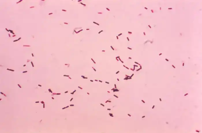
Gram staining protocols are among the most common microbiology lab methods. Gram staining helps differentiate many bacteria based on cell wall structure and staining response. It also gives information about cell shape and arrangement, such as cocci, rods, chains, clusters, or pairs.
NCBI Bookshelf explains that Gram staining differentiates bacterial species based on cell wall characteristics through a series of staining steps. It remains a widely used first-line method for bacterial classification and early laboratory assessment. See Gram Staining.
A useful Gram stain protocol should state the sample type, smear preparation, fixation approach, staining sequence, timing control, rinse method, microscope settings, positive and negative control organisms if used, and interpretation rules. It should also explain common errors, such as over-decolorization, under-decolorization, old cultures, thick smears, heat damage, mixed cultures, and poor microscope focusing.
Gram stain results should not be treated as a full identification by themselves. They are a useful clue, not a complete answer. Additional biochemical, molecular, culture, or clinical information may be needed depending on the purpose.
Microbial Growth Curves and OD600 Methods
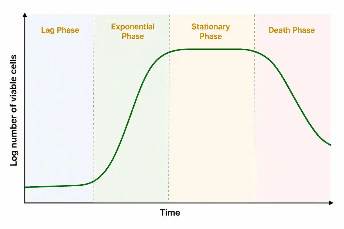
Microbial growth protocols measure how a population changes over time. Growth may be followed by optical density, viable plate counts, fluorescence, dry weight, metabolic signal, or automated growth curves.
OD600 is often used as a quick proxy for bacterial culture turbidity, but it is not a direct count of viable cells. It can be affected by organism shape, cell size, clumping, medium background, instrument path length, and whether cells are alive or dead. For quantitative work, OD600 often needs calibration against viable counts or another direct measurement.
A useful growth-curve protocol should state the organism, inoculum source, starting density, medium, vessel type, aeration, sampling interval, blank correction, replicate strategy, measurement wavelength, data transformation, and how growth phases or generation time will be estimated.
The major growth phases are usually described as lag phase, exponential or log phase, stationary phase, and death or decline phase. In practice, real curves can be messy because of nutrient limits, pH changes, oxygen availability, aggregation, evaporation, instrument differences, or stress responses.
Antimicrobial Susceptibility Testing Protocols
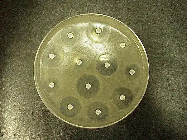
Antimicrobial susceptibility testing protocols help determine how a microorganism responds to antimicrobial agents under standardized conditions. These methods are important in clinical microbiology, public health, antimicrobial resistance monitoring, food safety, veterinary microbiology, and research.
Common methods include disk diffusion, broth dilution, broth microdilution, agar dilution, gradient diffusion, and automated systems. EUCAST explains that antimicrobial susceptibility testing is performed using phenotypic methods such as reference broth microdilution or standardized disk diffusion, and that minimum inhibitory concentration, or MIC, is the basis of susceptibility testing. See EUCAST Disk Diffusion and Quality Control.
ASM's Kirby-Bauer disk diffusion protocol describes disk diffusion as a standardized method and a practical alternative to broth dilution for laboratories without newer automated systems. See Kirby-Bauer Disk Diffusion Susceptibility Test Protocol.
A useful susceptibility testing protocol should state the organism, inoculum standardization method, medium, antimicrobial agent, disk or concentration range, incubation conditions, quality control strain if required, endpoint rule, zone or MIC interpretation system, and whether clinical breakpoints or research-only interpretation applies.
For public-facing educational content, be careful not to oversimplify susceptibility testing. Zone size or MIC only becomes meaningful when the method is standardized and interpreted using an accepted system such as EUCAST or CLSI guidance.
Biofilm Assays and Crystal Violet Staining
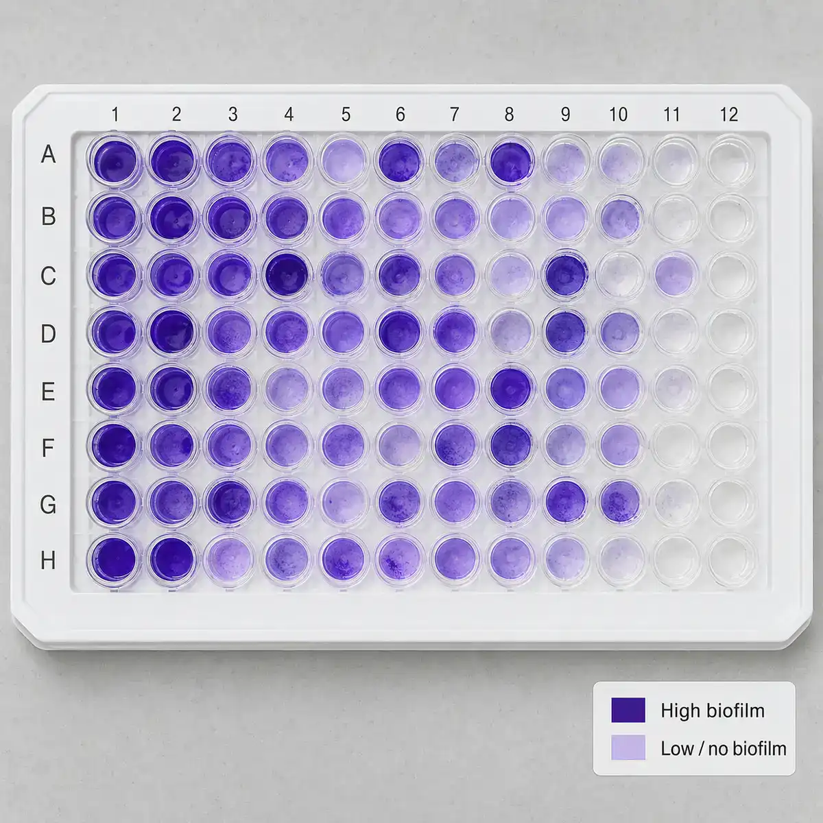
Biofilm protocols study microbial cells attached to surfaces and embedded in extracellular material. Biofilms matter in medicine, environmental microbiology, water systems, industrial fouling, dental microbiology, food processing, and antimicrobial resistance research.
A common biofilm method is the microtiter dish biofilm formation assay, often using crystal violet staining to estimate attached biomass. A widely cited JoVE protocol describes a microtiter dish assay for studying biofilm formation by Pseudomonas aeruginosa. See Microtiter Dish Biofilm Formation Assay. A review of biofilm assessment methods also discusses quantitative and qualitative approaches, including crystal violet assays. See Quantitative and Qualitative Assessment Methods for Biofilm Growth and Biofilm Formation.
A useful biofilm protocol should state the organism, surface, medium, incubation conditions, wash method, stain, solubilization method if used, plate reader settings, biomass interpretation, replicate strategy, and whether viable cells, total biomass, matrix material, or architecture are being measured.
Crystal violet is useful, but it does not directly measure live cells. It stains attached biomass. A treatment can reduce viability without removing biomass, or remove biomass without proving that every cell was killed. Good biofilm work often pairs biomass assays with viability, microscopy, CFU counts, or molecular methods.
Environmental, Food and Water Microbiology Protocols
Microbiology protocols are not limited to pure lab cultures. Environmental, food, and water microbiology often involve mixed communities, low-abundance organisms, stressed cells, complex matrices, inhibitory substances, and sampling bias.
A useful environmental or food microbiology protocol should define the sample source, sampling container, transport conditions, hold time, homogenization method, dilution strategy, selective or nonselective media, incubation conditions, target organism or indicator group, detection limit, and reporting unit.
For molecular environmental microbiology, microbial DNA extraction and PCR-based methods may reveal organisms that do not grow under the chosen culture conditions. For culture-based methods, colony recovery depends on the medium and conditions. Both approaches have strengths and limitations.
This is an area where future BioExplorer sub-pages can be especially useful: water testing methods, environmental swab protocols, soil microbial DNA extraction, food sample dilution, microbial indicator organisms, and culture-independent microbiome methods all deserve their own focused pages.
Microbial Molecular Methods
Microbial molecular biology protocols use DNA or RNA to study microorganisms. These methods include microbial DNA extraction, colony PCR, 16S rRNA gene amplification, qPCR, sequencing prep, metagenomics, strain typing, plasmid analysis, and detection of resistance genes.
A useful microbial DNA protocol should state the organism or sample type, lysis method, contamination controls, extraction blank, DNA quality check, PCR inhibition check, primer target, positive control, negative control, and downstream analysis. Environmental and low-biomass samples need special caution because contamination can become part of the result.
For broader molecular methods, see BioExplorer's Molecular Biology Methods and Protocols page, which covers DNA extraction, RNA isolation, PCR, qPCR, gel electrophoresis, cloning, sequencing preparation, and molecular troubleshooting.
How to Choose the Right Microbiology Protocol
Do not choose a microbiology protocol only because it appears first in search results. Choose it because it matches your organism, sample, safety level, research question, detection method, and data goal.
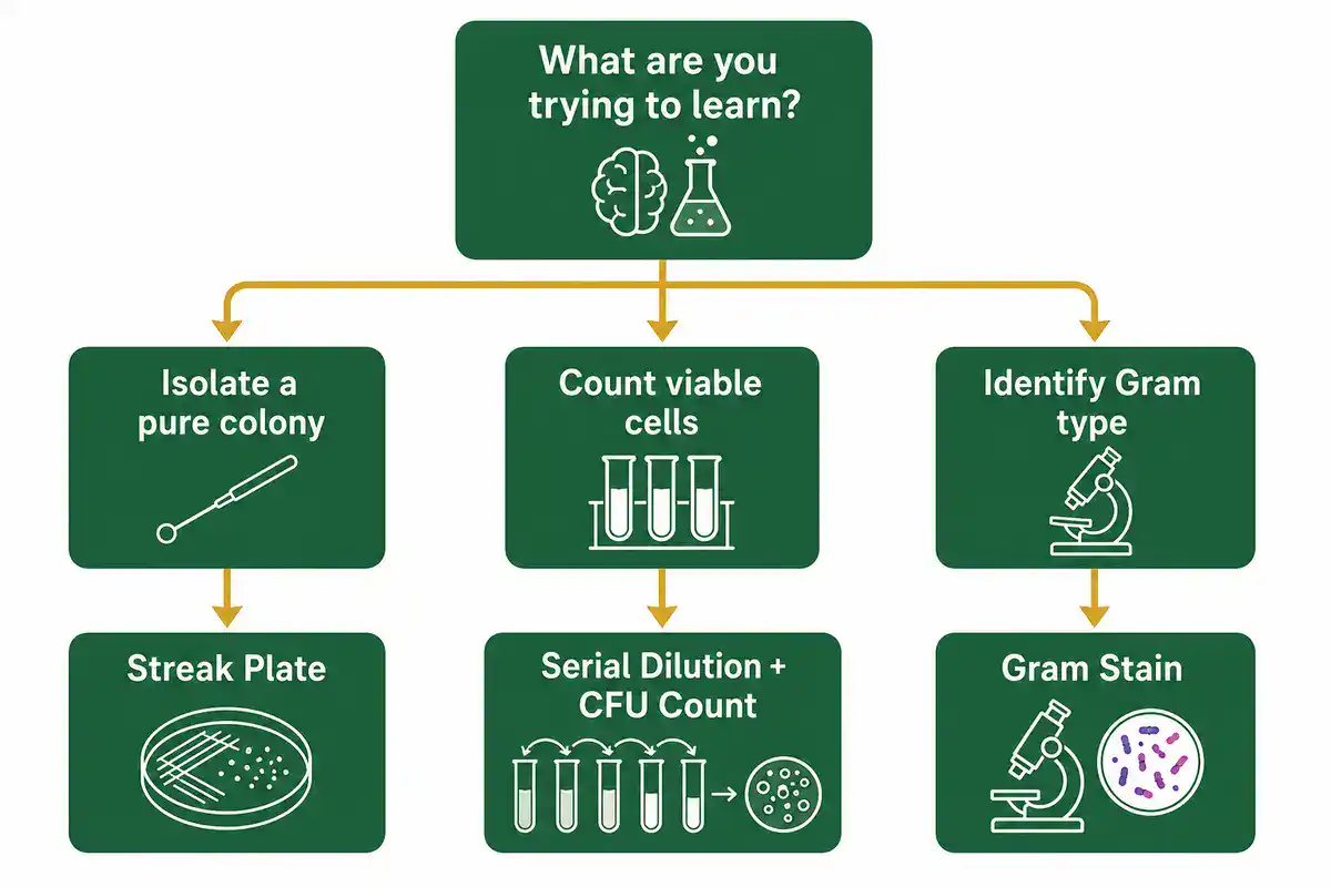
Before using any protocol, check these points:
- Organism or sample type: known strain, mixed culture, environmental sample, food sample, water sample, clinical specimen, fungus, bacterium, archaeon, virus, or unknown material.
- Goal: isolation, enumeration, identification, growth measurement, viability, antimicrobial response, contamination check, biofilm study, or molecular detection.
- Biosafety: risk group, biosafety level, PPE, engineering controls, aerosol risk, waste disposal, and institutional approval.
- Culture conditions: medium, oxygen, temperature range, incubation time, humidity, light, selective agents, and plate or broth format.
- Controls: positive control, negative control, sterile media control, extraction blank, no-template control, quality control strain, or untreated control.
- Readout: colony morphology, Gram stain, CFU/mL, OD600, zone diameter, MIC, PCR band, qPCR Ct value, sequencing result, or biofilm absorbance.
- Limitations: culturable organisms only, clumping, mixed cultures, viable but nonculturable cells, dead-cell DNA, contamination, inhibitors, or nonstandard incubation conditions.
- Verification: know how success will be checked before beginning the protocol.
Common Mistakes in Microbiology Protocols
Microbiology methods often fail for ordinary reasons. The method may be valid, but the culture, sample, dilution, controls, or safety assumptions may not fit the question.
- Skipping biosafety review: unknown samples and unfamiliar organisms should not be handled casually.
- Poor labeling: tubes, plates, dilutions, dates, organisms, and treatments must be traceable.
- Weak aseptic technique: contamination can create false growth, false positives, or mixed cultures.
- Using wet plates: excess surface moisture can make colonies spread and distort counts.
- Counting the wrong plate: plates with too few, too many, spreader, or merged colonies can give misleading CFU results.
- Forgetting dilution factors: CFU/mL calculations depend on dilution and plated volume.
- Overinterpreting Gram stains: staining gives useful clues, but it is not full species identification.
- Using old or stressed cultures: age and growth phase can affect staining, susceptibility testing, and physiology.
- Ignoring controls: sterile controls, known organisms, quality control strains, and no-template controls help distinguish real results from error.
- Assuming OD600 equals viable cell count: turbidity can include dead cells, clumps, debris, or non-growing cells.
- Comparing nonstandard susceptibility tests: antimicrobial results need standardized methods and accepted interpretation rules.
- Not recording deviations: small changes in medium, incubation, timing, or handling can explain major differences.
Microbiology Calculators and Lab Tools
Microbiology protocols often depend on calculations. A wrong dilution, plated volume, CFU conversion, log reduction, growth-rate estimate, or MIC interpretation can change the experiment before the results are even discussed.
Useful microbiology calculators include:
- serial dilution calculator
- CFU/mL calculator
- CFU/g calculator
- plate count calculator
- log reduction calculator
- dilution factor calculator
- OD600 dilution calculator
- bacterial growth rate calculator
- generation time calculator
- MIC dilution calculator
- zone of inhibition comparison tool
- biofilm biomass normalization calculator
- qPCR microbial load calculator
- contamination rate calculator
- plate layout calculator
BioExplorer's Biology Tools and Calculators hub is building free browser-based tools by branch of biology. The existing Genetics and Inheritance Tools section includes inheritance and population genetics tools, while the Botany Tools section supports plant science workflows. As BioExplorer expands its microbiology tools, this page can link directly to CFU calculators, dilution tools, growth-curve tools, antimicrobial testing calculators, and biofilm analysis helpers.
Trusted Microbiology Protocol Resources
Use BioExplorer as a guide, but always check your institution's approved SOPs, biosafety rules, manufacturer instructions, and supervisor guidance before performing real laboratory work. These external resources are useful starting points for microbiology protocols and method background:
- CDC Biosafety in Microbiological and Biomedical Laboratories: biosafety guidance based on protocol-driven risk assessment in biomedical and microbiological laboratories.
- CDC Biological Risk Assessment: guidance on identifying hazards, evaluating risk, applying controls, and reviewing control effectiveness.
- Aseptic Laboratory Techniques: Plating Methods: peer-reviewed microbiology education article covering streak plate, spread plate, and pour plate methods.
- ASM Serial Dilution Protocols: microbiology protocol resource for serial dilution and standard plate count concepts.
- NCBI Bookshelf: Gram Staining: background on Gram staining and bacterial differentiation by cell wall characteristics.
- Plate Counting You Can Count On: microbiology commentary on plate-counting logic and countable colony ranges.
- Bacterial Culture Through Selective and Non-Selective Conditions: review covering bacterial culture, solid media, colony morphology, and culture conditions.
- EUCAST Disk Diffusion and Quality Control: antimicrobial susceptibility testing methodology, disk diffusion, broth microdilution, MIC, and interpretation concepts.
- ASM Kirby-Bauer Disk Diffusion Protocol: standardized disk diffusion susceptibility testing protocol.
- Microtiter Dish Biofilm Formation Assay: peer-reviewed protocol article for a microtiter plate biofilm assay.
- Biofilm Assessment Methods Review: review of quantitative and qualitative biofilm assessment methods.
Safety and Responsibility
Microbiology protocols can involve living microorganisms, unknown samples, infectious material, aerosols, sharps, incubators, chemical disinfectants, stains, UV light, autoclaves, biosafety cabinets, centrifuges, and regulated waste. This page is educational. It does not replace formal training, institutional SOPs, biosafety approval, chemical safety guidance, or supervision by qualified personnel.
Do not culture or manipulate unknown organisms, clinical specimens, environmental isolates, recombinant organisms, or potentially infectious material without the proper facility, training, approval, containment, and disposal procedures. When in doubt, stop and follow institutional biosafety guidance.
Related BioExplorer Resources
- Biology Methods and Protocols: main hub for biology lab methods, protocol categories, safety notes, and trusted resources.
- Microbiology: guide to bacteria, archaea, fungi, protists, viruses, microbial ecology, and microbial research.
- Microbiology Glossary: clear definitions of bacteria, fungi, culture, contamination, biofilm, Gram stain, colony, and related microbial terms.
- Molecular Biology Methods and Protocols: protocols for DNA, RNA, PCR, cloning, gel electrophoresis, sequencing prep, and molecular analysis.
- Cell Biology Methods and Protocols: methods for cell culture, cell counting, viability, staining, microscopy, flow cytometry, and cell-based assays.
- Biochemistry Methods and Protocols: methods for protein assays, SDS-PAGE, western blotting, enzyme kinetics, chromatography, and protein purification.
- Biology Tools and Calculators: free browser-based biology calculators and educational tools.
Frequently Asked Questions
Microbiology protocols are written laboratory workflows for handling, culturing, staining, counting, identifying, or testing microorganisms under defined and safe conditions.
Common microbiology protocols include aseptic technique, media preparation, streak plating, spread plating, serial dilution, CFU counting, Gram staining, bacterial culture, microbial growth curves, antimicrobial susceptibility testing, and biofilm assays.
Aseptic technique reduces contamination and helps protect cultures, samples, workers, and the environment. Poor aseptic technique can create false results, mixed cultures, and unsafe working conditions.
CFU means colony-forming unit. It estimates viable microbial cells or cell groups that can form visible colonies under the chosen culture conditions.
No. OD600 measures culture turbidity, not direct viable cell number. It can be affected by dead cells, clumps, debris, organism shape, and instrument settings. Viable counts usually require plating or another viability method.
Gram staining helps differentiate many bacteria by cell wall staining behavior and gives clues about cell shape and arrangement. It is useful, but it is not complete species identification by itself.
Not always. Online protocols vary in quality and may not match your organism, biosafety level, equipment, sample type, or institution's rules. For real lab work, follow approved SOPs, biosafety guidance, manufacturer instructions, and supervisor training.
Cite this page
BioExplorer. (2026, July 11). Microbiology Protocols. https://www.bioexplorer.net/methods_and_protocols/microbiology/