
Biochemistry Methods and Protocols
Biochemistry protocols are practical laboratory methods used to study proteins, enzymes, metabolites, buffers, biochemical reactions, molecular interactions, and the chemical processes that make living systems work. These methods help researchers measure protein concentration, separate proteins by size, detect target proteins, test enzyme activity, purify biomolecules, prepare buffers, and connect biochemical data back to a biological question.
This page is a guide to major biochemistry methods and protocols used in research labs, teaching labs, biotechnology, molecular biology, cell biology, immunology, microbiology, and biomedical science. It explains what each method is for, what a good protocol should include, where mistakes usually happen, and which trusted resources can help you go deeper.
A useful protocol is not just a list of steps. It should explain the purpose of the method, the sample type, reagents, controls, equipment, timing, data readout, safety issues, troubleshooting, and limitations. Nature Protocols describes strong protocols as including reagents, equipment, timing, procedures, design advice, limitations, troubleshooting, data analysis, and result interpretation.
Biochemistry Methods and Protocols Guide:
- What Are Biochemistry Protocols?
- Biochemistry Protocols Guide
- Core Biochemistry Methods at a Glance
- Protein Extraction and Sample Preparation
- Protein Concentration Assay Protocols
- Bradford Assay
- BCA Assay
- SDS-PAGE and Protein Electrophoresis Protocols
- Western Blot Protocols
- Enzyme Assay Protocols
- Enzyme Kinetics, Km, Vmax and Inhibition
- Protein Purification and Chromatography Protocols
- Buffer Preparation and pH Control
- Spectrophotometry, Fluorescence and Standard Curves
- Mass Spectrometry and Proteomics Methods
- How to Choose the Right Biochemistry Protocol
- Common Mistakes in Biochemistry Protocols
- Biochemistry Calculators and Lab Tools
- Trusted Biochemistry Protocol Resources
- Safety and Responsibility
- Related BioExplorer Resources
- Frequently Asked Questions
What Are Biochemistry Protocols?
Biochemistry protocols are written workflows for studying biological molecules and chemical reactions inside living systems. Many focus on proteins because proteins act as enzymes, receptors, transporters, structural components, antibodies, channels, motors, and signaling molecules.
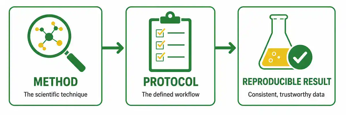
The method is the scientific technique. The protocol is the practical workflow for using that technique under defined conditions. For example, SDS-PAGE is a method for separating proteins by apparent molecular weight. An SDS-PAGE protocol describes the gel system, sample preparation, reducing or non-reducing conditions, ladder, running buffer, voltage, staining method, and interpretation of bands.
Biochemistry overlaps closely with molecular biology, cell biology, biotechnology, immunology, microbiology, pharmacology, physiology, structural biology, proteomics, and medicine.
Biochemistry Protocols Guide
Use this page as a map for common biochemistry lab methods and protein analysis workflows.
- protein extraction and sample preparation
- protein concentration assays
- Bradford, BCA, Lowry, and UV absorbance methods
- SDS-PAGE and native PAGE
- western blotting and antibody detection
- enzyme activity assays and enzyme kinetics
- Michaelis-Menten analysis, Km, Vmax, and kcat
- protein purification and chromatography
- buffer preparation and pH control
- spectrophotometry and fluorescence assays
- protein stability and interaction methods
- mass spectrometry and proteomics
- controls, troubleshooting, and quality checks
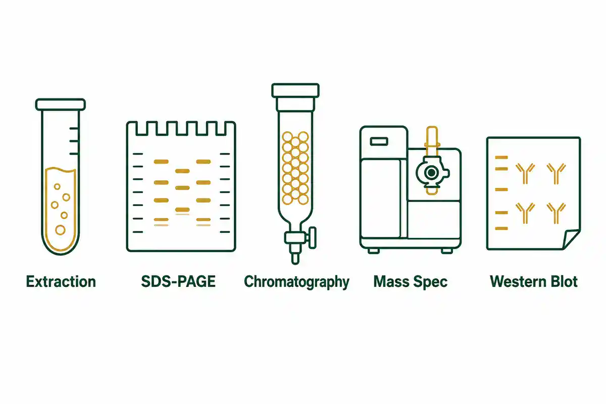
Core Biochemistry Methods at a Glance
The table below gives a quick map of important biochemistry methods and what each one helps measure.
| Method | Main Purpose | Common Readout |
|---|---|---|
| Protein extraction | Release proteins from cells, tissues, microbes, or organelles | Total protein lysate, soluble fraction, membrane fraction, clarified extract |
| Bradford assay | Estimate protein concentration using Coomassie dye binding | Absorbance near 595 nm compared with a standard curve |
| BCA assay | Estimate protein concentration using copper reduction and bicinchoninic acid | Absorbance near 562 nm compared with a standard curve |
| SDS-PAGE | Separate denatured proteins by apparent molecular weight | Protein bands on a stained gel or transferred membrane |
| Native PAGE | Separate proteins under non-denaturing conditions | Protein complexes, isoforms, or activity-retaining bands |
| Western blot | Detect a specific protein using antibodies | Target band size, signal intensity, background, loading control |
| Enzyme assay | Measure catalytic activity under defined conditions | Reaction rate, product formation, substrate loss, activity units |
| Enzyme kinetics | Analyze how reaction rate changes with substrate concentration | Km, Vmax, kcat, catalytic efficiency, inhibition pattern |
| Chromatography | Separate and purify proteins or metabolites | Fractions, peaks, purity, yield, activity, absorbance |
| Mass spectrometry | Identify proteins, peptides, modifications, or molecular masses | Mass-to-charge data, peptide spectra, protein IDs, proteomics results |
If you need structural, binding, or force based techniques such as X-ray crystallography, SPR, or AFM, see BioExplorer’s Biophysics Methods and Protocols guide.
Protein Extraction and Sample Preparation
Many biochemistry protocols begin with sample preparation. Before a protein can be measured, separated, purified, or detected, it usually has to be released from cells or tissues without destroying the feature being studied.
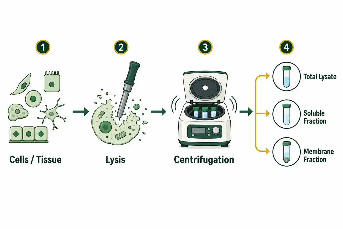
A protein extraction protocol should clearly state the sample type, lysis method, buffer composition, protease or phosphatase inhibitor use, temperature, centrifugation conditions, fraction collected, storage conditions, and downstream assay. A whole-cell lysate for western blotting is not the same as a native extract for enzyme activity, a membrane fraction for receptor analysis, or a purified protein preparation for structural work.
The key question is not only "how do I extract protein?" The better question is "what form of the protein do I need to preserve?" A protocol for enzyme activity may avoid harsh denaturants. A protocol for SDS-PAGE may intentionally denature proteins. A protocol for phosphorylation analysis may require phosphatase inhibitors. A protocol for membrane proteins may require detergents that do not destroy the assay.
Protein Concentration Assay Protocols
Protein concentration assays estimate how much protein is present in a sample. This matters before loading an SDS-PAGE gel, normalizing a western blot, comparing enzyme activity, preparing a purification step, or reporting protein yield.
Common protein assay methods include Bradford, BCA, Lowry, UV absorbance at 280 nm, and dye-binding or fluorescence-based assays. A good protein assay protocol should specify the standard used, sample dilution range, blank, replicates, wavelength, standard curve, compatible buffers, and known interfering substances.
Bradford Assay
The Bradford protein assay uses Coomassie dye to estimate total protein. Thermo Fisher explains that proteins bind Coomassie dye in acidic conditions, causing a spectral shift, and that 595 nm is commonly used to measure the blue dye-protein complex. See Bradford assay principles.
The Bradford assay is fast and widely used, but it is not automatically compatible with every sample. Detergents, very acidic conditions, protein-to-protein variation, and low molecular weight peptides can affect interpretation. If your protein sample contains detergents or unusual buffer components, check assay compatibility before trusting the number.
BCA Assay
The BCA protein assay is another common protein quantification method. Thermo Fisher's Pierce BCA guide explains that the method combines reduction of copper ions by protein in alkaline conditions with bicinchoninic acid detection, forming a purple complex with strong absorbance at 562 nm. See Pierce BCA Protein Assay Kit user guide.
BCA assays are useful for many protein samples, but reducing agents, chelators, detergents, and buffer components may interfere depending on the exact formulation. The safest approach is to run standards and unknowns in the same buffer when possible, keep the readings inside the linear range, and avoid extrapolating beyond the standard curve.
SDS-PAGE and Protein Electrophoresis Protocols
SDS-PAGE protocols are central to biochemistry because they separate proteins by apparent molecular weight. In SDS-PAGE, proteins are usually denatured and coated with sodium dodecyl sulfate, which gives them a more uniform negative charge relative to mass. When an electric field is applied, smaller proteins generally migrate farther through the gel matrix than larger proteins.
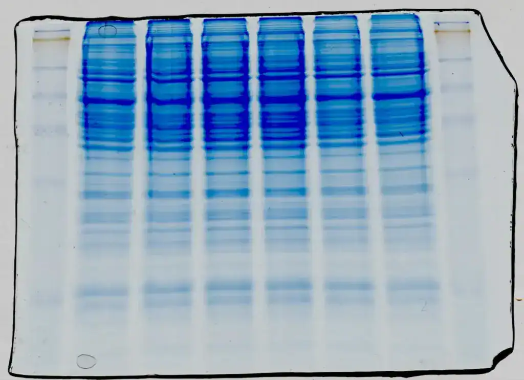
NCBI Bookshelf explains that SDS-PAGE separates proteins using a detergent and discontinuous buffer system, and that SDS gives denatured proteins a uniform charge-to-mass ratio. See Western Blot: Principles, Procedures, and Clinical Applications.
A useful SDS-PAGE protocol should describe sample buffer, reducing or non-reducing conditions, gel percentage, protein ladder, sample amount, running buffer, voltage, run time, stain, destain, imaging method, and interpretation. If the gel is being used before a western blot, the protocol should also connect the gel to transfer conditions and antibody detection.
Common SDS-PAGE problems include overloaded lanes, smiling bands, smeared bands, weak staining, poor separation, protein degradation, sample precipitation, and mismatched gel percentage. A good protocol helps readers understand these problems rather than only telling them to "run a gel."
Western Blot Protocols
Western blotting detects a specific protein from a complex sample using gel separation, membrane transfer, and antibody-based detection. It is widely used in biochemistry, cell biology, molecular biology, immunology, pharmacology, and biomedical research.
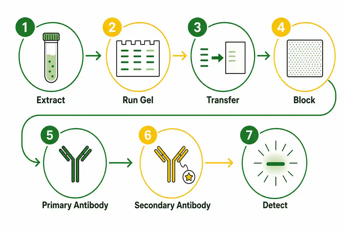
NCBI Bookshelf describes western blotting as a workflow in which electrophoresis separates proteins, proteins are transferred to a membrane, and specific proteins are detected. It also notes common troubleshooting issues such as multiple bands, high background, antibody concentration, transfer conditions, and membrane handling. See Western Blot: Principles, Procedures, and Clinical Applications.
A strong western blot protocol should include protein extraction conditions, quantification method, loading amount, gel system, transfer method, membrane type, blocking conditions, primary antibody, secondary antibody, washing, detection chemistry, exposure or imaging settings, loading control, positive control, negative control, and normalization method.
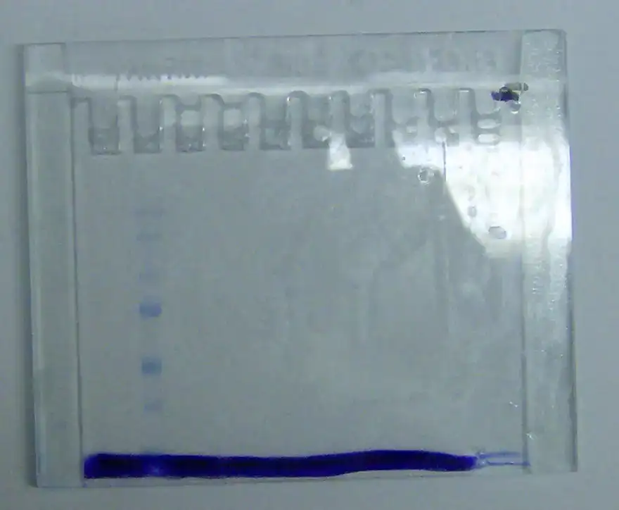
Western blots can be persuasive, but they are not self-interpreting. A band at the expected size supports target detection, but it does not prove specificity by itself. Antibody validation, controls, loading normalization, exposure range, replicate strategy, and sample preparation all matter.
Enzyme Assay Protocols
Enzyme assay protocols measure catalytic activity under defined conditions. They are used to study metabolism, signaling, drug inhibition, enzyme regulation, protein engineering, diagnostics, food science, biotechnology, and toxicology.
An enzyme assay may measure product formation, substrate disappearance, absorbance change, fluorescence change, luminescence, pH shift, oxygen consumption, or coupled reaction output. A useful enzyme assay protocol should state the enzyme source, substrate, buffer, pH, temperature, cofactors, detection method, time course, linear range, background control, blank, positive control, and unit definition.
NCBI's Assay Guidance Manual emphasizes that enzyme assays should establish initial velocity conditions and that substrate concentration can be varied to estimate Km and Vmax. It also notes that background can be measured by leaving out enzyme or substrate. See Basics of Enzymatic Assays for HTS.
The most common mistake in enzyme assays is treating a single endpoint as if it automatically represents enzyme activity. If the reaction is not linear with time and enzyme amount, the result may reflect substrate depletion, product inhibition, enzyme instability, detector saturation, or assay interference rather than true catalytic rate.
Enzyme Kinetics, Km, Vmax and Inhibition
Enzyme kinetics protocols ask how reaction rate changes with substrate concentration, enzyme concentration, inhibitors, pH, temperature, cofactors, or mutations. These protocols are more than ordinary activity assays because they are used to estimate kinetic parameters and compare enzyme behavior.
Important enzyme kinetics terms include:
- Initial velocity: the early reaction rate measured before substrate depletion or product accumulation changes the reaction.
- Km: the substrate concentration at which reaction velocity is half of Vmax under Michaelis-Menten assumptions.
- Vmax: the maximum reaction velocity under saturating substrate conditions.
- kcat: the turnover number, often used to describe the number of substrate molecules converted per enzyme active site per unit time.
- Catalytic efficiency: commonly expressed as kcat/Km for comparing enzyme performance under certain conditions.
- IC50: the inhibitor concentration that reduces signal or activity by 50 percent under a specific assay setup.
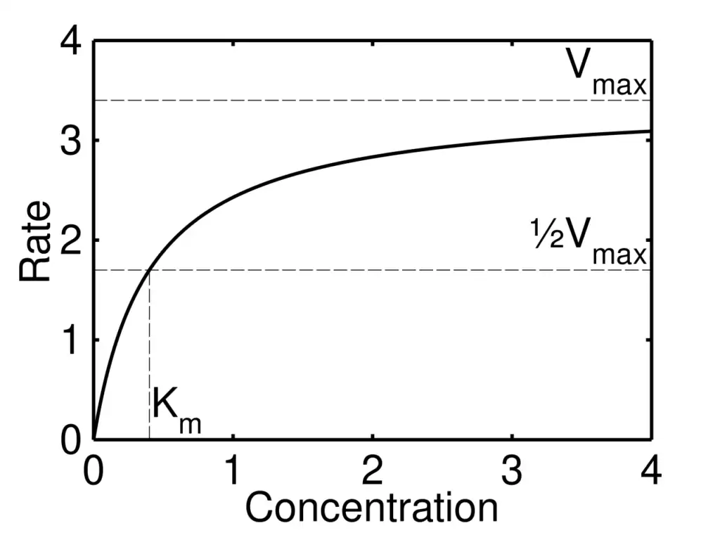
The Assay Guidance Manual explains that Km and Vmax should be estimated using initial velocity conditions and a saturation curve, and it cautions against traditional linearized methods when nonlinear regression is more appropriate. See Measurement of Km and Vmax.
For searchers looking for enzyme assay protocols, the key is this: a protocol should not only say what to pipette. It should explain how the signal relates to enzyme activity, what controls remove background, and whether the data actually support the kinetic model being used.
Protein Purification and Chromatography Protocols
Protein purification protocols separate a target protein from other proteins, nucleic acids, salts, metabolites, detergents, cell debris, and contaminants. Purification may be done for enzyme assays, antibody production, structural biology, binding studies, mass spectrometry, biochemical characterization, or biotechnology production.
Chromatography is one of the most important protein purification method families. Cytiva explains that purification success is often described by purity, homogeneity, and yield, and that chromatography methods can separate proteins by specific ligand recognition, metal ion binding, charge, size, hydrophobicity, or a combination of properties. See protein purification chromatography principles.
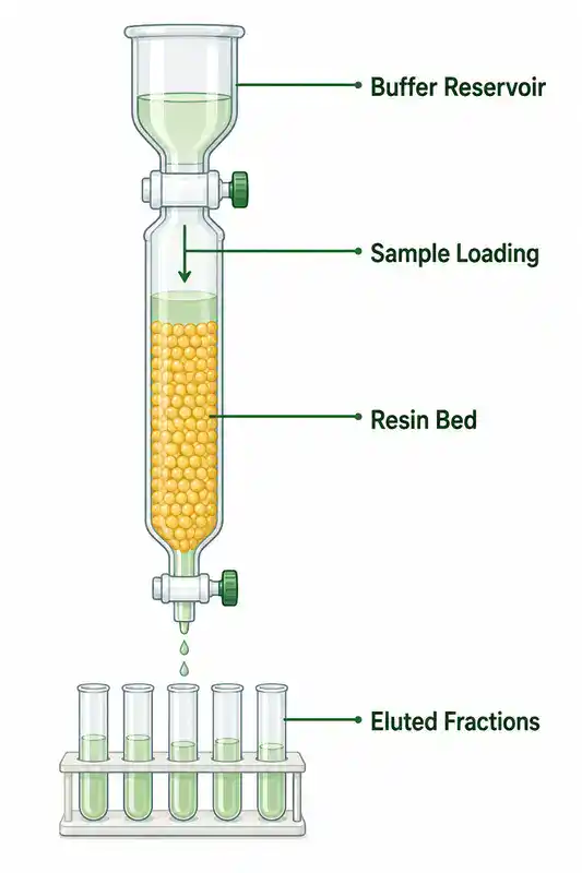
Common chromatography methods include:
- Affinity chromatography: separates proteins based on specific binding interactions, such as tagged proteins or antibody-antigen binding.
- IMAC: immobilized metal affinity chromatography, often used for histidine-tagged recombinant proteins.
- Ion exchange chromatography: separates proteins based on charge.
- Size exclusion chromatography: separates molecules based on size and is often used to assess monomers, aggregates, or complexes.
- Hydrophobic interaction chromatography: separates based on hydrophobic surface properties.
- Reverse phase chromatography: commonly used for peptides and analytical separations.
A useful protein purification protocol should track yield and purity across each step. The final protein may look clean on a gel but still be inactive, aggregated, degraded, contaminated, or unstable. For enzymes, activity recovery matters as much as band purity.
Buffer Preparation and pH Control
Biochemistry protocols depend heavily on buffers. Proteins and enzymes are sensitive to pH, salt concentration, temperature, reducing agents, metal ions, detergents, stabilizers, and preservatives. A buffer that works for one protein can destabilize another.
A good buffer protocol should state the buffer compound, target pH, temperature of pH adjustment, final concentration, salt concentration, additives, storage conditions, sterilization method if needed, and compatibility with downstream assays. Some buffers interfere with protein assays, enzyme assays, chromatography resins, or mass spectrometry.
Common buffer-related mistakes include adjusting pH before all components are dissolved, ignoring temperature effects on pH, using old reducing agents, adding incompatible detergents, forgetting chelators, and assuming that "same pH" means "same biochemical behavior."
Spectrophotometry, Fluorescence and Standard Curves
Many biochemistry methods rely on absorbance or fluorescence. A spectrophotometer or plate reader may be used for protein assays, enzyme assays, nucleic acid purity, metabolic assays, dye-binding methods, reporter assays, and kinetic measurements.
A useful spectrophotometry or fluorescence protocol should include wavelength, instrument path length or path length correction, blank, standard curve, linear range, replicate strategy, background subtraction, sample dilution, and how results will be calculated.
Standard curves are especially important. If the unknown sample is outside the standard curve range, the result should not be treated as reliable. Dilute and rerun the sample rather than forcing a number from a curve that does not cover it.
Mass Spectrometry and Proteomics Methods
Proteomics methods study many proteins at once. Mass spectrometry can help identify proteins, peptides, post-translational modifications, protein complexes, and changes in protein abundance across conditions.
NCBI Bookshelf explains that mass spectrometers measure mass, and that in proteomics, mass information can help reveal protein identity, chemical modifications, and structure. It also describes the basic mass spectrometer components: source, analyzer, and detector. See Mass Spectrometry for Proteomics.
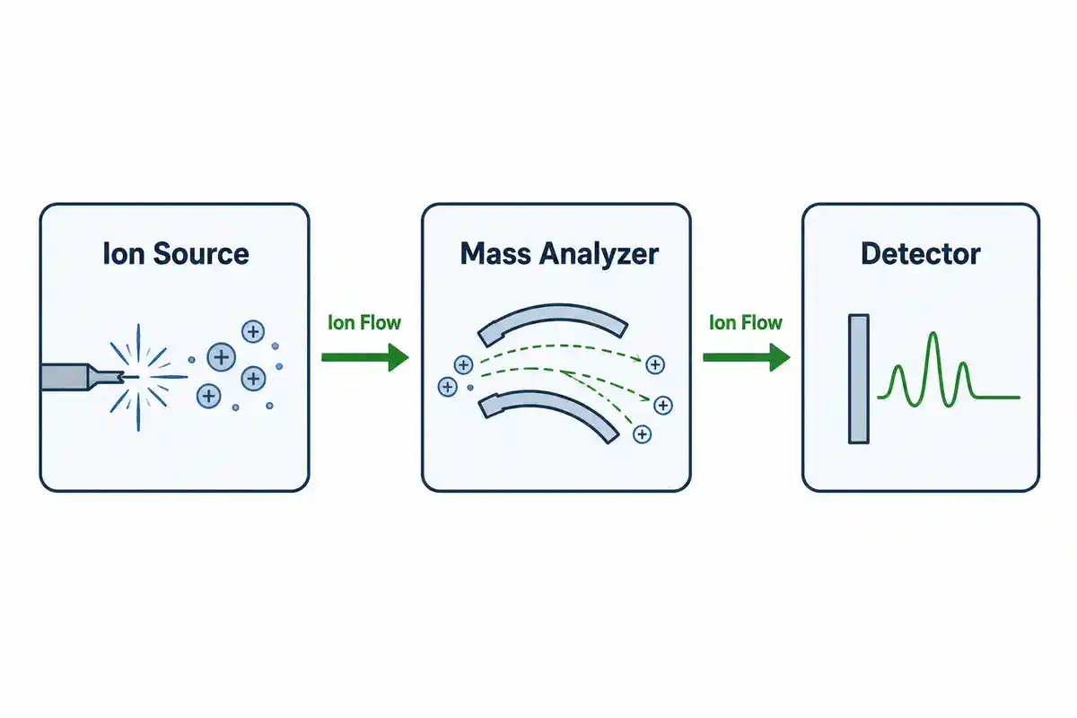
Proteomics protocols usually require careful sample cleanup, digestion, peptide handling, instrument quality control, database searching, false discovery control, normalization, and biological interpretation. This is why proteomics is not simply "advanced western blotting." It is a different workflow with different strengths, limits, and data analysis requirements.
How to Choose the Right Biochemistry Protocol
Do not choose a biochemistry protocol only because it appears first in search results. Choose it because it matches your sample, molecule, detection method, instrument, safety requirements, and biological question.
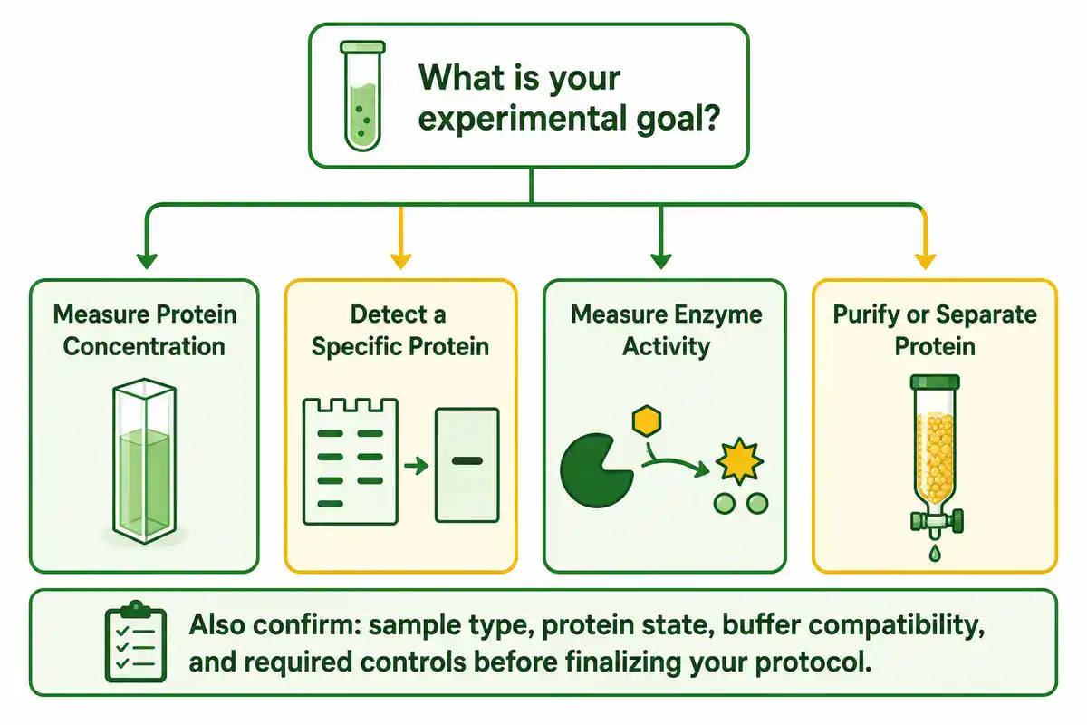
Before using any protocol, check these points:
- Sample type: purified protein, cell lysate, tissue lysate, serum, plasma, bacterial extract, plant extract, membrane fraction, organelle fraction, or enzyme preparation.
- Goal: concentration measurement, activity assay, protein detection, purification, binding analysis, inhibitor testing, stability testing, or proteomics.
- Protein state: native, denatured, reduced, oxidized, phosphorylated, glycosylated, membrane-bound, tagged, or complexed.
- Buffer compatibility: detergents, salts, reducing agents, chelators, glycerol, imidazole, urea, guanidine, and preservatives can affect assays.
- Controls: blanks, standards, positive controls, negative controls, loading controls, no-enzyme controls, no-substrate controls, and reference samples may be needed.
- Instrument settings: wavelength, gain, exposure, path length, voltage, transfer method, flow rate, column type, and detector settings should be documented.
- Safety: acrylamide, organic solvents, strong acids or bases, UV light, high voltage, biological samples, and some stains require appropriate safety handling.
- Verification: know how success will be checked before beginning the protocol.
Common Mistakes in Biochemistry Protocols
Biochemistry methods often fail for ordinary reasons. The assay may be valid, but the sample, buffer, timing, or detection range may not fit the protocol.
- Using the wrong protein assay: Bradford, BCA, Lowry, and UV methods do not have the same compatibility profile.
- Ignoring the standard curve: unknowns outside the standard curve range should be diluted and measured again.
- Overloading gels: too much protein can cause distorted SDS-PAGE bands and poor western blot transfer.
- Skipping loading controls: western blot signal is hard to interpret without normalization and proper controls.
- Using unvalidated antibodies: antibody specificity affects western blot interpretation.
- Measuring enzyme activity outside the linear range: endpoint signal may not represent true initial velocity.
- Forgetting background controls: enzyme assays often need no-enzyme, no-substrate, blank, or matrix controls.
- Changing pH or temperature without recording it: proteins and enzymes can respond strongly to small condition changes.
- Assuming purified means active: a protein can be pure but misfolded, degraded, aggregated, or inactive.
- Trusting a protocol without checking the source: peer-reviewed protocols, manufacturer instructions, institutional SOPs, and lab-validated workflows should carry more weight than copied web notes.
Biochemistry Calculators and Lab Tools
Biochemistry protocols often depend on calculations. A wrong dilution, protein concentration, buffer pH, enzyme unit conversion, gel loading amount, or standard curve calculation can damage the experiment before the assay begins.
Useful biochemistry calculators include:
- molarity calculator
- dilution calculator
- serial dilution calculator
- percent solution calculator
- buffer pH calculator
- protein concentration calculator
- Bradford assay calculator
- BCA assay calculator
- standard curve calculator
- enzyme activity calculator
- Michaelis-Menten calculator
- Km and Vmax calculator
- IC50 calculator
- SDS-PAGE loading calculator
- western blot antibody dilution calculator
BioExplorer's Biology Tools and Calculators hub is building free browser-based tools by branch of biology. The existing Genetics and Inheritance Tools section includes inheritance and population genetics tools, while the Botany Tools section supports plant science workflows. As BioExplorer expands its biochemistry tools, this page can link directly to protein assay calculators, enzyme kinetics calculators, buffer tools, and lab math calculators.
Trusted Biochemistry Protocol Resources
Use BioExplorer as a guide, but always check your institution's approved SOPs, safety rules, manufacturer instructions, and supervisor guidance before performing real laboratory work. These external resources are useful starting points for biochemistry protocols and method background:
- Nature Protocols: peer-reviewed protocol articles with design advice, troubleshooting, data analysis, limitations, and result interpretation.
- Bio-protocol: peer-reviewed open access protocol journal focused on improving research reproducibility.
- protocols.io: a platform for creating, versioning, sharing, and running reproducible methods.
- NCBI Bookshelf: Western Blot: background on SDS-PAGE, transfer, antibody detection, troubleshooting, and western blot safety.
- NCBI Assay Guidance Manual: Enzymatic Assays: guidance on enzyme assay design, background controls, initial velocity, Km, Vmax, and inhibition studies.
- Thermo Fisher Bradford Assay Principles: Coomassie dye-binding chemistry, wavelength guidance, and assay limitations.
- Thermo Fisher Pierce BCA Protein Assay Guide: BCA assay chemistry, absorbance at 562 nm, working range, standards, and compatibility notes.
- Cytiva Chromatography Principles: protein purification principles, chromatography method selection, purity, homogeneity, and yield.
- NCBI Bookshelf: Mass Spectrometry for Proteomics: background on mass spectrometry, proteomics, ionization, analyzers, and detectors.
- CDC Biosafety in Microbiological and Biomedical Laboratories: biosafety guidance for protocol-driven risk assessment in biomedical and microbiological laboratories.
Safety and Responsibility
Biochemistry protocols can involve biological samples, acrylamide, strong acids or bases, detergents, organic solvents, high voltage electrophoresis systems, UV light, centrifuges, sharps, stains, antibodies, enzymes, and specialized instruments. This page is educational. It does not replace formal training, institutional SOPs, chemical safety guidance, biosafety approval, or supervision by qualified personnel.
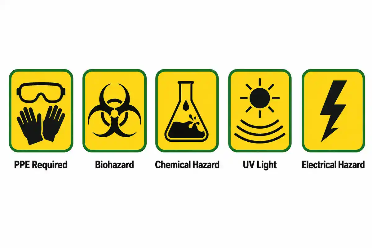
The CDC's biosafety guidance emphasizes protocol-driven risk assessment because no single document can identify every possible combination of risks and mitigation steps in biomedical laboratories. For any protocol involving human material, infectious agents, recombinant organisms, hazardous chemicals, or regulated samples, use approved institutional procedures and safety oversight.
Related BioExplorer Resources
- Biology Methods and Protocols: main hub for biology lab methods, protocol categories, safety notes, and trusted resources.
- Biochemistry: overview of biochemistry as the study of chemical processes in living systems.
- Biochemistry Glossary: clear definitions of enzymes, proteins, amino acids, ATP, metabolism, buffers, pH, and related biochemical terms.
- Molecular Biology: DNA, RNA, proteins, gene expression, molecular mechanisms, and related research methods.
- Molecular Biology Methods and Protocols: protocols for DNA, RNA, PCR, cloning, gel electrophoresis, sequencing prep, and molecular analysis.
- Cell Biology: guide to cells, organelles, cell signaling, cell division, and cell-based experiments.
- Biology Tools and Calculators: free browser-based biology calculators and educational tools.
Frequently Asked Questions
Biochemistry protocols are written laboratory workflows for studying proteins, enzymes, metabolites, biochemical reactions, buffers, molecular interactions, and chemical processes in living systems.
Common biochemistry protocols include protein extraction, protein concentration assays, Bradford assay, BCA assay, SDS-PAGE, western blotting, enzyme activity assays, enzyme kinetics, chromatography, buffer preparation, and protein purification.
SDS-PAGE separates proteins by apparent molecular weight. Western blotting usually uses SDS-PAGE first, then transfers the separated proteins to a membrane and detects a specific target protein with antibodies.
The best protein assay depends on your sample, buffer, detergent, reducing agent, protein type, expected concentration, instrument, and downstream use. Bradford and BCA assays are common, but each has compatibility limits.
Controls help separate true enzyme activity from background signal, substrate instability, reagent interference, detector noise, or non-enzymatic reaction. Useful controls may include no-enzyme, no-substrate, blank, positive control, and inhibitor control conditions.
Km is the substrate concentration at which the reaction velocity is half of Vmax under Michaelis-Menten assumptions. It is useful for comparing enzyme behavior, but it must be measured under suitable initial velocity conditions.
Not always. Online protocols vary in quality and may not match your sample, equipment, reagents, safety level, or institution's rules. For real lab work, follow approved SOPs, manufacturer instructions, safety guidance, and supervisor instruction.
Cite this page
BioExplorer. (2026, September 30). Biochemistry Methods and Protocols. https://www.bioexplorer.net/methods_and_protocols/biochemistry/